• 文献检索
  • 文档翻译
  • 深度研究
  • 学术资讯
  • Suppr Zotero 插件Zotero 插件
  • 邀请有礼
  • 套餐&价格
  • 历史记录
应用&插件
Suppr Zotero 插件Zotero 插件浏览器插件Mac 客户端Windows 客户端微信小程序
定价
高级版会员购买积分包购买API积分包
服务
文献检索文档翻译深度研究API 文档MCP 服务
关于我们
关于 Suppr公司介绍联系我们用户协议隐私条款
关注我们

Suppr 超能文献

核心技术专利:CN118964589B侵权必究
粤ICP备2023148730 号-1Suppr @ 2026

文献检索

告别复杂PubMed语法,用中文像聊天一样搜索,搜遍4000万医学文献。AI智能推荐,让科研检索更轻松。

立即免费搜索

文件翻译

保留排版,准确专业,支持PDF/Word/PPT等文件格式,支持 12+语言互译。

免费翻译文档

深度研究

AI帮你快速写综述,25分钟生成高质量综述,智能提取关键信息,辅助科研写作。

立即免费体验

自体骨填充的手术性骨缺损中的骨再生:大鼠的荧光显微镜分析。

Bone regeneration in surgically created defects filled with autogenous bone: an epifluorescence microscopy analysis in rats.

机构信息

Discipline of Oral and Maxillofacial Surgery, Department of Surgery and Integrated Clinics, Araçatuba Dental School, São Paulo State University, Araçatuba, SP, Brazil.

出版信息

J Appl Oral Sci. 2010 Jul-Aug;18(4):346-53. doi: 10.1590/s1678-77572010000400005.

DOI:10.1590/s1678-77572010000400005
PMID:20835568
原文链接:https://pmc.ncbi.nlm.nih.gov/articles/PMC5349066/
Abstract

UNLABELLED

Although the search for the ideal bone substitute has been the focus of a large number of studies, autogenous bone is still the gold standard for the filling of defects caused by pathologies and traumas, and mainly, for alveolar ridge reconstruction, allowing the titanium implants installation.

OBJECTIVES

The aim of this study was to evaluate the dynamics of autogenous bone graft incorporation process to surgically created defects in rat calvaria, using epifluorescence microscopy.

MATERIAL AND METHODS

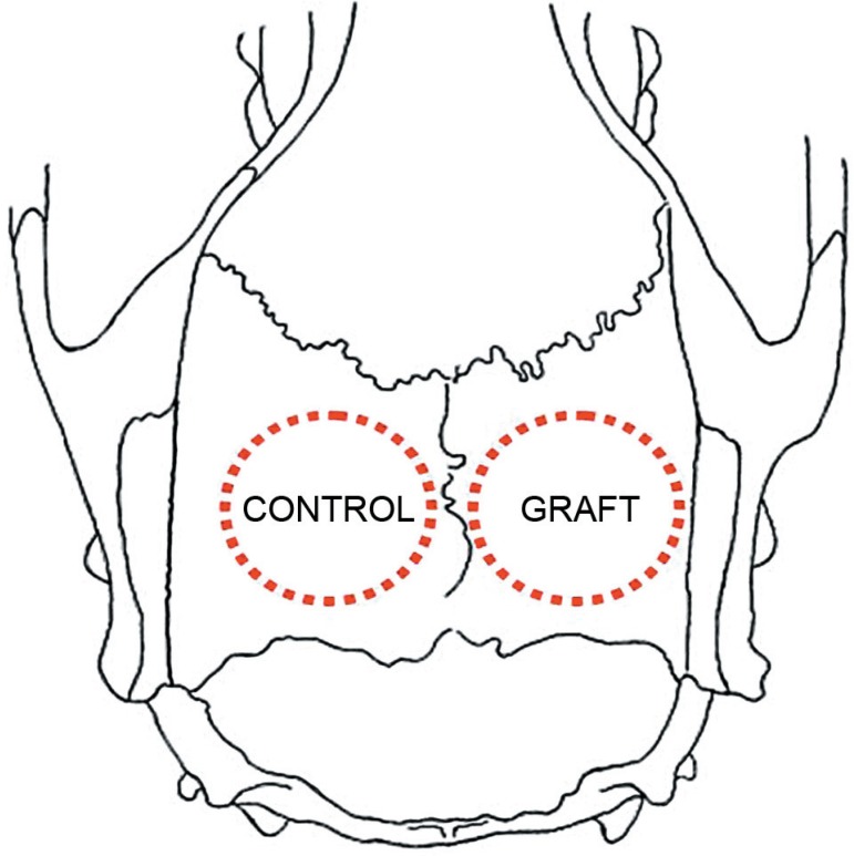
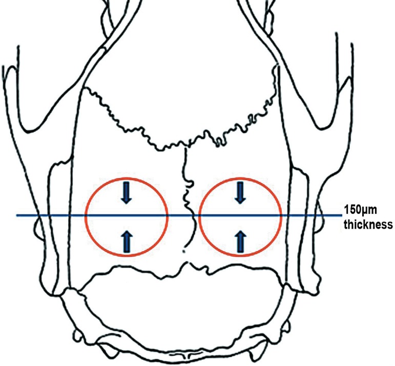
Five adult male rats weighing 200-300 g were used. The animals received two 5-mm-diameter bone defects bilaterally in each parietal bone with a trephine bur under general anesthesia. Two groups of defects were formed: a control group (n=5), in which the defects were filled with blood clot, and a graft group (n=5), in which the defects were filled with autogenous bone block, removed from the contralateral defect. The fluorochromes calcein and alizarin were applied at the 7th and 30th postoperative days, respectively. The animals were killed at 35 days.

RESULTS

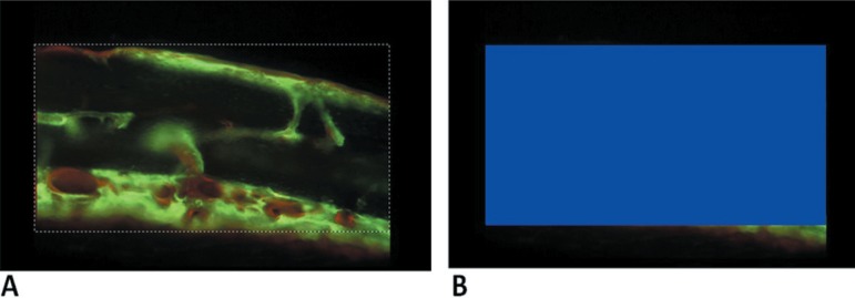
The mineralization process was more intense in the graft group (32.09%) and occurred mainly between 7 and 30 days, the period labeled by calcein (24.66%).

CONCLUSIONS

The fluorochromes showed to be appropriate to label mineralization areas. The interfacial areas between fluorochrome labels are important sources of information about the bone regeneration dynamics.

摘要

未加标签

尽管人们一直在寻找理想的骨替代物,但自体骨仍然是填充因疾病和创伤引起的缺陷的金标准,主要用于牙槽嵴重建,以允许钛植入物的安装。

目的

本研究旨在使用荧光显微镜评估自体骨移植物在大鼠颅骨手术性缺损中的吸收过程的动力学。

材料和方法

使用 5 只成年雄性大鼠,体重 200-300g。在全身麻醉下,用环钻在每块顶骨上双侧形成两个 5mm 直径的骨缺损。形成两组缺陷:对照组(n=5),其中缺陷用血凝块填充;移植物组(n=5),其中缺陷用取自对侧缺陷的自体骨块填充。在术后第 7 天和第 30 天分别应用钙黄绿素和茜素红两种荧光染料。在第 35 天处死动物。

结果

移植物组的矿化过程更强烈(32.09%),主要发生在第 7 天至第 30 天之间,即钙黄绿素标记的时期(24.66%)。

结论

荧光染料适用于标记矿化区域。荧光染料标记的界面区域是骨再生动力学的重要信息来源。

https://cdn.ncbi.nlm.nih.gov/pmc/blobs/f054/5349066/434823ecbec7/jaos-18-04-0346-g12.jpg
https://cdn.ncbi.nlm.nih.gov/pmc/blobs/f054/5349066/f7b060d462ce/jaos-18-04-0346-g01.jpg
https://cdn.ncbi.nlm.nih.gov/pmc/blobs/f054/5349066/e422588c8c3e/jaos-18-04-0346-g02.jpg
https://cdn.ncbi.nlm.nih.gov/pmc/blobs/f054/5349066/189a7d914b06/jaos-18-04-0346-g03.jpg
https://cdn.ncbi.nlm.nih.gov/pmc/blobs/f054/5349066/e9be590c3343/jaos-18-04-0346-g04.jpg
https://cdn.ncbi.nlm.nih.gov/pmc/blobs/f054/5349066/0b861d41b7ac/jaos-18-04-0346-g05.jpg
https://cdn.ncbi.nlm.nih.gov/pmc/blobs/f054/5349066/1cce0a746726/jaos-18-04-0346-g06.jpg
https://cdn.ncbi.nlm.nih.gov/pmc/blobs/f054/5349066/da1d748af9df/jaos-18-04-0346-g07.jpg
https://cdn.ncbi.nlm.nih.gov/pmc/blobs/f054/5349066/c6918cd324f0/jaos-18-04-0346-g08.jpg
https://cdn.ncbi.nlm.nih.gov/pmc/blobs/f054/5349066/20895b05c05a/jaos-18-04-0346-g09.jpg
https://cdn.ncbi.nlm.nih.gov/pmc/blobs/f054/5349066/75339ff8aff2/jaos-18-04-0346-g10.jpg
https://cdn.ncbi.nlm.nih.gov/pmc/blobs/f054/5349066/289593de8623/jaos-18-04-0346-g11.jpg
https://cdn.ncbi.nlm.nih.gov/pmc/blobs/f054/5349066/434823ecbec7/jaos-18-04-0346-g12.jpg

相似文献

1
Bone regeneration in surgically created defects filled with autogenous bone: an epifluorescence microscopy analysis in rats.自体骨填充的手术性骨缺损中的骨再生:大鼠的荧光显微镜分析。
J Appl Oral Sci. 2010 Jul-Aug;18(4):346-53. doi: 10.1590/s1678-77572010000400005.
2
The use of hydroxyapatite and autogenous cancellous bone grafts to repair bone defects in rats.使用羟基磷灰石和自体松质骨移植修复大鼠骨缺损。
Int J Oral Maxillofac Surg. 2005 Mar;34(2):178-84. doi: 10.1016/j.ijom.2004.06.005.
3
Evaluation of the presence of VEGF, BMP2 and CBFA1 proteins in autogenous bone graft: histometric and immunohistochemical analysis.自体骨移植中血管内皮生长因子、骨形态发生蛋白2和核心结合因子α1蛋白的存在情况评估:组织计量学和免疫组织化学分析。
J Craniomaxillofac Surg. 2014 Jun;42(4):333-9. doi: 10.1016/j.jcms.2013.05.022. Epub 2013 Aug 7.
4
Allogeneic bone grafting of calvarial defects: an experimental study in the rabbit.颅骨缺损的同种异体骨移植:在兔身上的实验研究。
Int J Oral Maxillofac Surg. 2002 Oct;31(5):525-31. doi: 10.1054/ijom.2002.0281.
5
Repair process of surgical defects filled with autogenous bone grafts in tibiae of diabetic rats.糖尿病大鼠胫骨中填充自体骨移植的手术缺损修复过程。
J Appl Oral Sci. 2008 Sep-Oct;16(5):316-20. doi: 10.1590/s1678-77572008000500003.
6
Early effect of platelet-rich plasma on bone healing in combination with an osteoconductive material in rat cranial defects.富血小板血浆联合骨传导材料对大鼠颅骨缺损骨愈合的早期影响。
Clin Oral Implants Res. 2007 Apr;18(2):244-51. doi: 10.1111/j.1600-0501.2006.01327.x.
7
The effect of low-level laser on bone healing in critical size defects treated with or without autogenous bone graft: an experimental study in rat calvaria.低强度激光对采用或未采用自体骨移植治疗的临界尺寸骨缺损愈合的影响:大鼠颅骨的实验研究
Clin Oral Implants Res. 2014 Oct;25(10):1131-6. doi: 10.1111/clr.12239. Epub 2013 Aug 6.
8
Evaluation of the long-term results of rat cranial bone repair using a particular xenograft.使用特定异种移植物评估大鼠颅骨修复的长期结果。
J Oral Implantol. 2010;36(3):167-73. doi: 10.1563/AAID-JOI-D-09-00064.
9
Effect of LLLT on autogenous bone grafts in the repair of critical size defects in the calvaria of immunosuppressed rats.低强度激光疗法对免疫抑制大鼠颅骨临界尺寸缺损修复中自体骨移植的影响。
J Craniomaxillofac Surg. 2014 Oct;42(7):1196-202. doi: 10.1016/j.jcms.2014.02.008. Epub 2014 Feb 25.
10
Bony healing of large cranial and mandibular defects protected from soft-tissue interposition: A comparative study of spontaneous bone regeneration, osteoconduction, and cancellous autografting in dogs.防止软组织介入的大型颅骨和下颌骨缺损的骨愈合:犬自发性骨再生、骨传导和松质骨自体移植的比较研究
Plast Reconstr Surg. 1998 Mar;101(3):660-72. doi: 10.1097/00006534-199803000-00013.

引用本文的文献

1
Preparation of Collagen/Hydroxyapatite Composites Using the Alternate Immersion Method and Evaluation of the Cranial Bone-Forming Capability of Composites Complexed with Acidic Gelatin and b-FGF.采用交替浸泡法制备胶原蛋白/羟基磷灰石复合材料并评估与酸性明胶和碱性成纤维细胞生长因子复合的复合材料的颅骨形成能力。
Materials (Basel). 2022 Dec 9;15(24):8802. doi: 10.3390/ma15248802.
2
Vertical Guided Bone Regeneration in the Rabbit Calvarium Using Porous Nanohydroxyapatite Block Grafts Coated with rhVEGF and Cortical Perforation.多孔纳米羟基磷灰石块移植体联合 rhVEGF 涂层和皮质骨穿孔引导兔颅骨骨再生的实验研究
Int J Nanomedicine. 2020 Dec 10;15:10059-10073. doi: 10.2147/IJN.S268182. eCollection 2020.
3

本文引用的文献

1
Repair process of surgical defects filled with autogenous bone grafts in tibiae of diabetic rats.糖尿病大鼠胫骨中填充自体骨移植的手术缺损修复过程。
J Appl Oral Sci. 2008 Sep-Oct;16(5):316-20. doi: 10.1590/s1678-77572008000500003.
2
Bone regeneration after demineralized bone matrix and castor oil (Ricinus communis) polyurethane implantation.脱矿骨基质与蓖麻油(蓖麻)聚氨酯植入后的骨再生
J Appl Oral Sci. 2008 Mar-Apr;16(2):122-6. doi: 10.1590/s1678-77572008000200008.
3
The monocortical critical size bone defect as an alternative experimental model in testing bone substitute materials.
Dental stem cell sources and their potentials for bone tissue engineering.
牙源性干细胞来源及其在骨组织工程中的潜力。
J Istanb Univ Fac Dent. 2015 Apr 29;49(2):51-56. doi: 10.17096/jiufd.42908. eCollection 2015.
4
"Over-inlay" block graft and differential morphometry: a novel block graft model to study bone regeneration and host-to-graft interfaces in rats.“过度嵌体”块状移植与差异形态测量:一种用于研究大鼠骨再生及宿主与移植体界面的新型块状移植模型
J Periodontal Implant Sci. 2016 Aug;46(4):220-33. doi: 10.5051/jpis.2016.46.4.220. Epub 2016 Aug 30.
5
Comparison of the Effect of Velvet Antler from Different Sections on Longitudinal Bone Growth of Adolescent Rats.不同部位鹿茸对青春期大鼠纵向骨生长影响的比较
Evid Based Complement Alternat Med. 2016;2016:1927534. doi: 10.1155/2016/1927534. Epub 2016 Jun 13.
6
Effect of collagen sponge and fibrin glue on bone repair.胶原海绵和纤维蛋白胶对骨修复的影响。
J Appl Oral Sci. 2015 Nov-Dec;23(6):623-8. doi: 10.1590/1678-775720150374.
7
Deciphering the elusive nature of sharp bone trauma using epifluorescence macroscopy: a comparison study multiplexing classical imaging approaches.运用荧光显微镜技术解析隐匿性锐骨创伤的本质:多模式经典成像方法的对比研究。
Int J Legal Med. 2013 Jan;127(1):169-76. doi: 10.1007/s00414-012-0678-6. Epub 2012 Feb 23.
单皮质临界尺寸骨缺损作为测试骨替代材料的一种替代实验模型。
Oral Surg Oral Med Oral Pathol Oral Radiol Endod. 2006 Jul;102(1):7-13. doi: 10.1016/j.tripleo.2005.09.011. Epub 2006 Mar 24.
4
Sinus graft with biogran, autogenous bone, and PRP: a report of three cases with histology and micro-CT.使用生物颗粒、自体骨和富血小板血浆的鼻窦移植:三例组织学和显微CT报告
Int J Periodontics Restorative Dent. 2006 Apr;26(2):113-25.
5
Autogenous bone grafts and bone substitutes--tools and techniques: I. A 20,000-case experience in maxillofacial and craniofacial surgery.自体骨移植与骨替代物——工具与技术:I. 20000例颌面及颅面外科手术经验
Plast Reconstr Surg. 2005 Oct;116(5 Suppl):6S-24S. doi: 10.1097/01.prs.0000173862.20563.12.
6
Does platelet-rich plasma promote remodeling of autologous bone grafts used for augmentation of the maxillary sinus floor?富含血小板血浆是否能促进用于上颌窦底提升的自体骨移植的重塑?
Clin Oral Implants Res. 2005 Jun;16(3):349-56. doi: 10.1111/j.1600-0501.2005.01115.x.
7
Influence of implant microstructure on the dynamics of bone healing around immediate implants placed into periodontally infected sites. A confocal laser scanning microscopic study.种植体微观结构对植入牙周感染部位的即刻种植体周围骨愈合动力学的影响。一项共聚焦激光扫描显微镜研究。
Clin Oral Implants Res. 2004 Feb;15(1):44-53. doi: 10.1111/j.1600-0501.2004.00995.x.
8
Systemic effects on bone healing of a new hyaluronic acid-like bacterial exopolysaccharide.一种新型透明质酸样细菌胞外多糖对骨愈合的全身影响。
Calcif Tissue Int. 2003 Sep;73(3):232-6. doi: 10.1007/s00223-002-2081-7.
9
[The principles of tissue engineering: role of growth factors in the bone regeneration].[组织工程学原理:生长因子在骨再生中的作用]
Minerva Stomatol. 2002 Sep;51(9):351-9.
10
High concentrations of bioactive glass material (BioGran) vs. autogenous bone for sinus floor elevation.高浓度生物活性玻璃材料(BioGran)与自体骨用于上颌窦底提升的比较
Clin Oral Implants Res. 2002 Aug;13(4):428-36. doi: 10.1034/j.1600-0501.2002.130412.x.