Iwasaki Laura R, Crosby Michael J, Gonzalez Yoly, McCall Willard D, Marx David B, Ohrbach Richard, Nickel Jeffrey C
University of Missouri Kansas City, School of Dentistry, Departments of Orthodontics and Dentofacial Orthopedics, and Oral Biology, Kansas City, MO.
Orthop Rev (Pavia). 2009;1(2):90-93. doi: 10.4081/or.2009.e29.
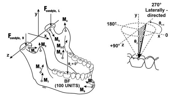
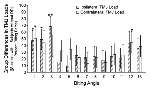
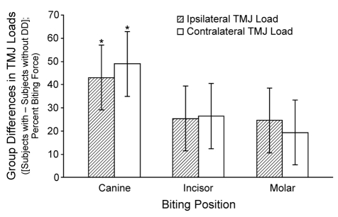
The likelihood of development of degenerative joint disease (DJD) of the temporomandibular joint (TMJ) is related to the integrity of the TMJ disc. Predilection for mechanical failure of the TMJ disc may reflect inter-individual differences in TMJ loads. Nine females and eight males in each of normal TMJ disc position and bilateral disc displacement diagnostic groups consented to participate in our study. Disc position was determined by bilateral magnetic resonance images of the joints. Three-dimensional (3D) anatomical geometry of each subject was used in a validated computer-assisted numerical model to calculate ipsilateral and contralateral TMJ loads for a range of biting positions (incisor, canine, molar) and angles (1-13). Each TMJ load was a resultant vector at the anterosuperi or-most mediolateral midpoint the condyle and characterized in terms of magnitude and 3D orientation. Analysis of variance (ANOVA) was used to test for effects of biting position and angle on TMJ loads. Mean TMJ loads in subjects with disc displacement were 9.5-69% higher than in subjects with normal disc position. During canine biting, TMJ loads in subjects with disc displacement were 43% (ipsilateral condyle, p=0.029) and 49% (contralateral condyle, p=0.015) higher on average than in subjects with normal disc position. Biting angle effects showed that laterally directed forces on the dentition produced ipsilateral joint loads, which on average were 69% higher (p=0.002) compared to individuals with normal TMJ disc position. The data reported here describe large differences in TMJ loads between individuals with disc displacement and normal disc position. The results support future investigations of inter-individual differences in joint mechanics as a variable in the development of DJD of the TMJ.
颞下颌关节(TMJ)退行性关节病(DJD)的发生可能性与TMJ盘的完整性有关。TMJ盘机械故障的易发性可能反映了TMJ负荷的个体差异。正常TMJ盘位置和双侧盘移位诊断组中各有9名女性和8名男性同意参与我们的研究。通过关节的双侧磁共振图像确定盘位置。在经过验证的计算机辅助数值模型中,使用每个受试者的三维(3D)解剖几何结构来计算一系列咬合位置(切牙、尖牙、磨牙)和角度(1 - 13)下同侧和对侧TMJ的负荷。每个TMJ负荷是髁突前上最中外侧中点处的合力矢量,并根据大小和3D方向进行表征。采用方差分析(ANOVA)来测试咬合位置和角度对TMJ负荷的影响。盘移位受试者的平均TMJ负荷比盘位置正常的受试者高9.5 - 69%。在尖牙咬合期间,盘移位受试者的TMJ负荷平均比盘位置正常的受试者高43%(同侧髁突,p = 0.029)和49%(对侧髁突,p = 0.015)。咬合角度效应表明,牙列上的侧向力会产生同侧关节负荷,与TMJ盘位置正常的个体相比,平均高出69%(p = 0.002)。此处报告的数据描述了盘移位个体和盘位置正常个体之间TMJ负荷的巨大差异。这些结果支持未来将关节力学个体差异作为TMJ的DJD发展中的一个变量进行研究。