• 文献检索
  • 文档翻译
  • 深度研究
  • 学术资讯
  • Suppr Zotero 插件Zotero 插件
  • 邀请有礼
  • 套餐&价格
  • 历史记录
应用&插件
Suppr Zotero 插件Zotero 插件浏览器插件Mac 客户端Windows 客户端微信小程序
定价
高级版会员购买积分包购买API积分包
服务
文献检索文档翻译深度研究API 文档MCP 服务
关于我们
关于 Suppr公司介绍联系我们用户协议隐私条款
关注我们

Suppr 超能文献

核心技术专利:CN118964589B侵权必究
粤ICP备2023148730 号-1Suppr @ 2026

文献检索

告别复杂PubMed语法,用中文像聊天一样搜索,搜遍4000万医学文献。AI智能推荐,让科研检索更轻松。

立即免费搜索

文件翻译

保留排版,准确专业,支持PDF/Word/PPT等文件格式,支持 12+语言互译。

免费翻译文档

深度研究

AI帮你快速写综述,25分钟生成高质量综述,智能提取关键信息,辅助科研写作。

立即免费体验

共焦扫描激光眼底镜在青光眼诊断与治疗中的应用

Confocal scanning laser ophthalmoscopy in glaucoma diagnosis and management.

作者信息

Alexandrescu C, Dascalu A M, Panca A, Sescioreanu A, Mitulescu C, Ciuluvica R, Voinea L, Celea C

机构信息

Carol Davila University of Medicine and Pharmacy, Bucharest, Romania.

出版信息

J Med Life. 2010 Jul-Sep;3(3):229-34.

PMID:20945812
原文链接:https://pmc.ncbi.nlm.nih.gov/articles/PMC3018995/
Abstract

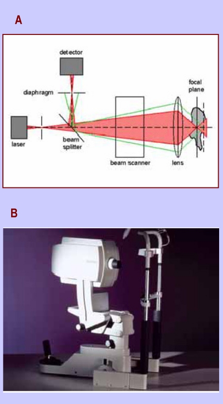
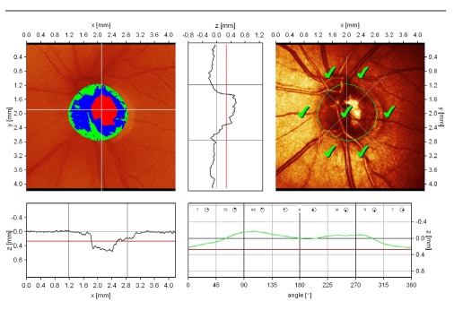
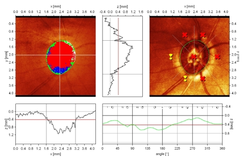
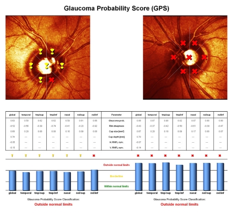
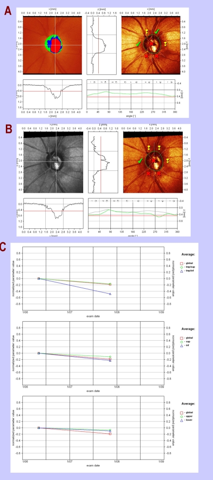
The early diagnosis and detection of progression are two key-elements in the actual management of glaucoma. The current opinion in clinical practice is to quantify the structural damage for a better follow-up of the patient and the standardization of the results. The present review is a concise survey of literature covering the period of 1990-2010, documenting the evidence-based role of confocal scanning laser ophthalmoscopy in glaucoma diagnosis and management.

摘要

青光眼实际管理中的两个关键要素是早期诊断和病情进展的检测。临床实践中的当前观点是量化结构损伤,以便更好地随访患者并使结果标准化。本综述是对1990年至2010年期间文献的简要调查,记录了共焦扫描激光眼科显微镜在青光眼诊断和管理中基于证据的作用。

https://cdn.ncbi.nlm.nih.gov/pmc/blobs/2776/3018995/71580a306184/JMedLife-03-229-g008.jpg
https://cdn.ncbi.nlm.nih.gov/pmc/blobs/2776/3018995/d17a626a8fef/JMedLife-03-229-g001.jpg
https://cdn.ncbi.nlm.nih.gov/pmc/blobs/2776/3018995/d10fd8db3160/JMedLife-03-229-g002.jpg
https://cdn.ncbi.nlm.nih.gov/pmc/blobs/2776/3018995/8765e0c5c71f/JMedLife-03-229-g003.jpg
https://cdn.ncbi.nlm.nih.gov/pmc/blobs/2776/3018995/2a202e729df7/JMedLife-03-229-g004.jpg
https://cdn.ncbi.nlm.nih.gov/pmc/blobs/2776/3018995/1f7dc0691cf8/JMedLife-03-229-g005.jpg
https://cdn.ncbi.nlm.nih.gov/pmc/blobs/2776/3018995/2e217a4a832b/JMedLife-03-229-g006.jpg
https://cdn.ncbi.nlm.nih.gov/pmc/blobs/2776/3018995/abb36a9cb976/JMedLife-03-229-g007.jpg
https://cdn.ncbi.nlm.nih.gov/pmc/blobs/2776/3018995/71580a306184/JMedLife-03-229-g008.jpg

相似文献

1
Confocal scanning laser ophthalmoscopy in glaucoma diagnosis and management.共焦扫描激光眼底镜在青光眼诊断与治疗中的应用
J Med Life. 2010 Jul-Sep;3(3):229-34.
2
[Comparative analysis of several morphometric parameters received using optic coherent tomography and scanning laser ophthalmoscopy in initial glaucoma diagnosis].
Vestn Oftalmol. 2011 Mar-Apr;127(2):18-20.
3
The effect of compression on clinical diagnosis of glaucoma based on non-analyzed confocal scanning laser ophthalmoscopy images.
Ophthalmic Surg Lasers Imaging. 2006 May-Jun;37(3):263; author reply 263-4. doi: 10.3928/15428877-20060501-17.
4
Comparing optic nerve head analysis between confocal scanning laser ophthalmoscopy and spectral domain optical coherence tomography.共焦扫描激光眼底镜与光谱域光学相干断层扫描在视神经乳头分析中的比较。
Curr Eye Res. 2014 Oct;39(10):1026-32. doi: 10.3109/02713683.2014.891752. Epub 2014 Mar 21.
5
Optic disc diameter measurement using planimeter and scanning laser ophthalmoscope.
Invest Ophthalmol Vis Sci. 1995 Oct;36(11):2162-3.
6
Prospective evaluation of optic nerve head by confocal scanning laser ophthalmoscopy after intraocular pressure control in adult glaucoma.成人青光眼眼压控制后通过共焦扫描激光眼底镜对视神经乳头进行前瞻性评估。
Semin Ophthalmol. 2013 Jan;28(1):13-8. doi: 10.3109/08820538.2012.730095.
7
Imaging of the optic disk in caring for patients with glaucoma: ophthalmoscopy and photography remain the gold standard.在青光眼患者的护理中,对视盘进行成像:检眼镜检查和照相仍然是金标准。
Surv Ophthalmol. 2014 Jul-Aug;59(4):454-8. doi: 10.1016/j.survophthal.2013.10.004. Epub 2013 Oct 16.
8
Can Visual Field Progression be Predicted by Confocal Scanning Laser Ophthalmoscopic Imaging of the Optic Nerve Head in Glaucoma? (An American Ophthalmological Society Thesis).青光眼患者视神经乳头的共焦扫描激光眼底镜成像能否预测视野进展?(美国眼科学会论文)
Trans Am Ophthalmol Soc. 2015;113:T4.
9
Correlations between digital planimetry and optical coherence tomography, confocal scanning laser ophthalmoscopy in assessment of optic disc parameters.数字测径术与光学相干断层扫描、共焦激光扫描检眼镜评估视盘参数的相关性。
Medicina (Kaunas). 2012;48(3):150-8.
10
The effects of study design and spectrum bias on the evaluation of diagnostic accuracy of confocal scanning laser ophthalmoscopy in glaucoma.研究设计和谱偏倚对共焦扫描激光眼科显微镜在青光眼诊断准确性评估中的影响。
Invest Ophthalmol Vis Sci. 2007 Jan;48(1):214-22. doi: 10.1167/iovs.06-0618.

引用本文的文献

1
Chemical Insights into Topical Agents in Intraocular Pressure Management: From Glaucoma Etiopathology to Therapeutic Approaches.眼压管理中局部用药的化学见解:从青光眼病因病理学到治疗方法
Pharmaceutics. 2024 Feb 15;16(2):274. doi: 10.3390/pharmaceutics16020274.
2
Three-Dimensional Light Field Fundus Imaging: Automatic Determination of Diagnostically Relevant Optic Nerve Head Parameters.三维眼底激光成像:自动确定有诊断意义的视盘参数。
Transl Vis Sci Technol. 2023 Jul 3;12(7):21. doi: 10.1167/tvst.12.7.21.
3
Macular Optical Coherence Tomography Imaging in Glaucoma.

本文引用的文献

1
The Heidelberg retina tomograph Glaucoma Probability Score: reproducibility and measurement of progression.海德堡视网膜断层扫描仪青光眼概率评分:可重复性和进展测量。
Ophthalmology. 2010 Apr;117(4):724-9. doi: 10.1016/j.ophtha.2009.09.036. Epub 2010 Jan 4.
2
HRT III glaucoma probability score and Moorfields regression across the glaucoma spectrum.HRT III青光眼概率评分与青光眼全谱的摩尔菲尔德回归分析
J Glaucoma. 2009 Jun-Jul;18(5):368-72. doi: 10.1097/IJG.0b013e31818c6edd.
3
Optic disc progression in glaucoma: comparison of confocal scanning laser tomography to optic disc photographs in a prospective study.
青光眼的黄斑光学相干断层扫描成像
J Ophthalmic Vis Res. 2021 Jul 29;16(3):478-489. doi: 10.18502/jovr.v16i3.9442. eCollection 2021 Jul-Sep.
4
Noninvasive monitoring of suprachoroidal, subretinal, and intravitreal implants using confocal scanning laser ophthalmoscope (cSLO) and optical coherence tomography (OCT).利用共焦扫描激光检眼镜(cSLO)和光相干断层扫描(OCT)对脉络膜上腔、视网膜下腔和眼内植入物进行无创监测。
Int J Pharm. 2021 Sep 5;606:120887. doi: 10.1016/j.ijpharm.2021.120887. Epub 2021 Jul 14.
5
Effect of color information on the diagnostic performance of glaucoma in deep learning using few fundus images.利用少量眼底图像的深度学习中颜色信息对青光眼诊断性能的影响
Int Ophthalmol. 2020 Nov;40(11):3013-3022. doi: 10.1007/s10792-020-01485-3. Epub 2020 Jun 27.
6
Macular imaging with optical coherence tomography in glaucoma.光学相干断层扫描在青光眼中的黄斑成像。
Surv Ophthalmol. 2020 Nov-Dec;65(6):597-638. doi: 10.1016/j.survophthal.2020.03.002. Epub 2020 Mar 19.
7
Medical Management of Glaucoma in the 21st Century from a Canadian Perspective.从加拿大视角看21世纪青光眼的医学管理
J Ophthalmol. 2016;2016:6509809. doi: 10.1155/2016/6509809. Epub 2016 Nov 8.
8
Repeatability of in vivo 3D lamina cribrosa microarchitecture using adaptive optics spectral domain optical coherence tomography.使用自适应光学光谱域光学相干断层扫描技术对体内三维筛板微结构的可重复性研究
Biomed Opt Express. 2014 Mar 10;5(4):1114-23. doi: 10.1364/BOE.5.001114. eCollection 2014 Apr 1.
9
Reflectance speckle of retinal nerve fiber layer reveals axonal activity.视网膜神经纤维层的反射斑显示轴突活动。
Invest Ophthalmol Vis Sci. 2013 Apr 12;54(4):2616-23. doi: 10.1167/iovs.12-11347.
青光眼视盘进展:前瞻性研究中激光共焦扫描断层成像与视盘照片的比较
Invest Ophthalmol Vis Sci. 2009 Apr;50(4):1682-91. doi: 10.1167/iovs.08-2457. Epub 2008 Dec 5.
4
Diagnostic tools for glaucoma detection and management.用于青光眼检测与管理的诊断工具。
Surv Ophthalmol. 2008 Nov;53 Suppl1(SUPPL1):S17-32. doi: 10.1016/j.survophthal.2008.08.003.
5
Comparison of HRT-3 glaucoma probability score and subjective stereophotograph assessment for prediction of progression in glaucoma.HRT-3青光眼概率评分与主观立体照片评估对青光眼病情进展预测的比较
Invest Ophthalmol Vis Sci. 2008 May;49(5):1898-906. doi: 10.1167/iovs.07-0111.
6
Diagnostic ability of Heidelberg Retina Tomograph 3 classifications: glaucoma probability score versus Moorfields regression analysis.海德堡视网膜断层扫描仪3分类的诊断能力:青光眼概率评分与摩尔菲尔德回归分析对比
Ophthalmology. 2007 Nov;114(11):1981-7. doi: 10.1016/j.ophtha.2007.01.015. Epub 2007 Apr 19.
7
Comparison of retinal nerve fibre layer thickness and visual field loss between different glaucoma groups.不同青光眼组视网膜神经纤维层厚度与视野缺损的比较。
Br J Ophthalmol. 2005 Aug;89(8):1004-7. doi: 10.1136/bjo.2004.050906.
8
Tomographic identification of neuroretinal rim loss in high-pressure, normal-pressure, and suspected glaucoma.
Invest Ophthalmol Vis Sci. 2004 Jul;45(7):2279-85. doi: 10.1167/iovs.03-1243.
9
Confocal scanning laser ophthalmoscopy classifiers and stereophotograph evaluation for prediction of visual field abnormalities in glaucoma-suspect eyes.共焦扫描激光检眼镜分类器及立体照片评估对可疑青光眼患者视野异常的预测作用
Invest Ophthalmol Vis Sci. 2004 Jul;45(7):2255-62. doi: 10.1167/iovs.03-1087.
10
Approach for identifying glaucomatous optic nerve progression by scanning laser tomography.
Invest Ophthalmol Vis Sci. 2003 Jun;44(6):2621-6. doi: 10.1167/iovs.02-0850.