Suppr超能文献

在晚期非小细胞肺癌中进行 CT 肿瘤体积测量:新兴临床工具的性能特征。

CT tumor volume measurement in advanced non-small-cell lung cancer: Performance characteristics of an emerging clinical tool.

机构信息

Department of Imaging, Dana-Farber Cancer Institute, Brigham and Women's Hospital, Boston, MA 02115, USA.

出版信息

Acad Radiol. 2011 Jan;18(1):54-62. doi: 10.1016/j.acra.2010.08.021. Epub 2010 Oct 30.

Abstract

RATIONALE AND OBJECTIVES

Determine inter- and intraobserver variability of computed tomography (CT) tumor volume measurements in advanced non-small-cell lung cancer (NSCLC) patients treated in a Phase II clinical trial using chest CT.

MATERIALS AND METHODS

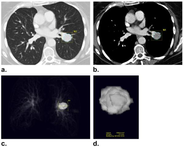
Twenty-three advanced NSCLC patients with a total of 53 measurable lung lesions enrolled in a Phase II, multicenter, open-label clinical trial of erlotinib were retrospectively studied with institutional review board approval. Two radiologists independently measured the tumor size, volume, and CT attenuation coefficient using commercially available volume analysis software. Concordance correlation coefficients (CCCs) and Bland-Altman plots were used to assess inter- and intraobserver agreement.

RESULTS

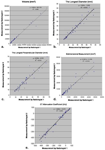
High CCCs (0.949-0.990) were observed in all types of measurements for interobserver agreement. The 95% limits of agreements for volume, unidimensional, and bidimensional measurements were (-26.0%, 18.6%), (-23.1%, 24.4%), and (-34.0%, 48.6%), respectively. Volume measurement had slightly higher CCC and narrower 95% limits of agreement compared to uni- and bidimensional measurements. CCCs for intraobserver agreement were high (range, 0.946-0.996) with CCC for volume being slightly higher than CCCs of uni- and bidimensional measurements. The smaller the tumor volume was, the larger the interobserver difference of CT attenuation. Location, morphology, or adjacent atelectasis had no significant impact on inter- or intraobserver variability.

CONCLUSION

CT tumor volume measurement in advanced NSCLC patients using clinical chest CT and commercially available software demonstrated high inter- and intraobserver agreement, indicating that the method may be used routinely in clinical practice.

摘要

背景与目的

采用胸部 CT 对接受 II 期临床试验治疗的晚期非小细胞肺癌(NSCLC)患者进行 CT 肿瘤体积测量,评估其观察者内和观察者间的可变性。

材料与方法

回顾性分析了 23 例接受厄洛替尼治疗的晚期 NSCLC 患者的 53 个可测量的肺部病变,这些患者均入组了一项多中心、开放标签的 II 期临床试验,该研究获得了机构审查委员会的批准。两名放射科医生使用商业可用的容积分析软件,分别独立地测量肿瘤的大小、体积和 CT 衰减系数。一致性相关系数(CCC)和 Bland-Altman 图用于评估观察者内和观察者间的一致性。

结果

在所有观察者间的测量中,均观察到了较高的 CCC(0.949-0.990)。体积、单维及二维测量的 95%一致性界限分别为(-26.0%,18.6%)、(-23.1%,24.4%)和(-34.0%,48.6%)。与单维和二维测量相比,体积测量的 CCC 略高,95%一致性界限也较窄。观察者内的一致性也很高(范围为 0.946-0.996),其中体积的 CCC 略高于单维和二维测量的 CCC。肿瘤体积越小,CT 衰减的观察者间差异越大。位置、形态或相邻的肺不张对观察者内和观察者间的变异性没有显著影响。

结论

使用临床胸部 CT 和商业上可用的软件对晚期 NSCLC 患者进行 CT 肿瘤体积测量,结果显示出了较高的观察者内和观察者间的一致性,这表明该方法可在临床实践中常规使用。

相似文献

1
CT tumor volume measurement in advanced non-small-cell lung cancer: Performance characteristics of an emerging clinical tool.
Acad Radiol. 2011 Jan;18(1):54-62. doi: 10.1016/j.acra.2010.08.021. Epub 2010 Oct 30.
5
Cone-beam CT-based delineation of stereotactic lung targets: the influence of image modality and target size on interobserver variability.
Int J Radiat Oncol Biol Phys. 2012 Feb 1;82(2):e265-72. doi: 10.1016/j.ijrobp.2011.03.042. Epub 2011 May 27.
6
Area--a consistent method to evaluate pulmonary tumor size on multidetector CT imaging: an intraobserver and interobserver agreement study.
Clin Imaging. 2013 Nov-Dec;37(6):1006-10. doi: 10.1016/j.clinimag.2013.06.011. Epub 2013 Aug 29.

引用本文的文献

1
Response Assessment of Primary Liver Tumors to Novel Therapies: an Imaging Perspective.
J Gastrointest Surg. 2023 Oct;27(10):2245-2259. doi: 10.1007/s11605-023-05762-1. Epub 2023 Jul 18.
2
A multi-modality physical phantom for mimicking tumor heterogeneity patterns in PET/CT and PET/MRI.
Med Phys. 2022 Sep;49(9):5819-5829. doi: 10.1002/mp.15853. Epub 2022 Jul 25.
4
Tumor volume dynamics and tumor growth rate in -rearranged advanced non-small-cell lung cancer treated with crizotinib.
Eur J Radiol Open. 2020 Jan 28;7:100210. doi: 10.1016/j.ejro.2019.12.004. eCollection 2020.
5
Pseudoprogression and hyperprogression in lung cancer: a comprehensive review of literature.
J Cancer Res Clin Oncol. 2020 Dec;146(12):3269-3279. doi: 10.1007/s00432-020-03360-1. Epub 2020 Aug 28.

本文引用的文献

2
Cancer statistics, 2009.
CA Cancer J Clin. 2009 Jul-Aug;59(4):225-49. doi: 10.3322/caac.20006. Epub 2009 May 27.
3
Pulmonary nodules: volume repeatability at multidetector CT lung cancer screening.
Radiology. 2009 Jun;251(3):919-25. doi: 10.1148/radiol.2513081313. Epub 2009 Apr 20.
4
Noncalcified lung nodules: volumetric assessment with thoracic CT.
Radiology. 2009 Apr;251(1):26-37. doi: 10.1148/radiol.2511071897.
8
Lung cancer: computerized quantification of tumor response--initial results.
Radiology. 2006 Dec;241(3):892-8. doi: 10.1148/radiol.2413051887.
9
Pulmonary nodules detected at lung cancer screening: interobserver variability of semiautomated volume measurements.
Radiology. 2006 Oct;241(1):251-7. doi: 10.1148/radiol.2411050860. Epub 2006 Aug 14.
10
Pulmonary metastases: effect of CT section thickness on measurement--initial experience.
Radiology. 2005 Mar;234(3):934-9. doi: 10.1148/radiol.2343040020. Epub 2005 Jan 28.

文献AI研究员

20分钟写一篇综述,助力文献阅读效率提升50倍。

立即体验

用中文搜PubMed

大模型驱动的PubMed中文搜索引擎

马上搜索

文档翻译

学术文献翻译模型,支持多种主流文档格式。

立即体验