Section of Minimally Invasive Surgery, Department of Surgery, Washington University School of Medicine, 660 South Euclid Avenue, Campus Box #8109, St. Louis, MO 63110, USA.
Surg Endosc. 2011 May;25(5):1390-4. doi: 10.1007/s00464-010-1373-7. Epub 2010 Nov 3.
The purpose of this study was to examine the biological environment of the esophageal hiatus through analysis of the collagen content within the gastrohepatic ligament (GHL), gastrophrenic ligament (GPL), and phrenoesophageal ligament (PEL) in patients with type I hiatal hernias (HH) and type III paraesophageal hernias (PEH).
A control group (N=10) and patients with type I HH (N=10) and type III PEH (N=10) were included in the analysis. Specimens of the GHL, PEL, and GPL were collected intraoperatively. Slides stained with sirius red/fast green were created and ten photos at 400×magnification were taken of each specimen. Axiovision 4.7 (Zeiss) photo analysis software was employed for quantification of collagen I (red) and III (green) by calculating color area (μm2). Statistical significance (p<0.05) was determined using a one-way ANOVA and Fisher's LSD post-test.
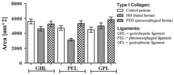
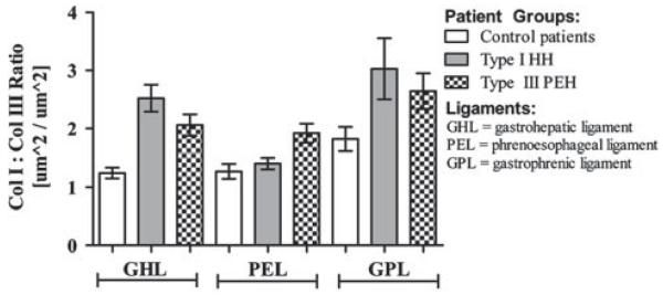
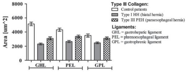
Cross-polarization microscopy revealed that the collagen I content was similar in the three study groups for the GHL, greater in the type III PEH group and in the control group compared to the type I HH group for the PEL, and greater in the type III PEH group compared to control group for the GPL. Collagen III quantity was greater in the control group than in the type I HH group for each ligament, and greater in the GHL and PEL when compared to the type III PEH group. Type III PEH patients had greater collagen III quantity than did type I HH patients for each ligament. Collagen type I:III ratio of the GHL was greater in both hernia groups compared to the control group. Type III PEH patients contained a higher I:III ratio than both the control and type I HH groups with respect to the PEL. There was no difference in the ratio with evaluation of the GPL for the three groups.
Evaluation of the esophageal hiatus revealed that patients with PEH have a different biological environment with regard to collagen content compared to control patients. The collagen I:III ratio of the study groups was equal to or greater than the control group. Collagen deficiency in the GE junction supporting ligaments does not appear to be an etiology of PEH formation.
本研究旨在通过分析Ⅰ型食管裂孔疝(HH)和Ⅲ型食管旁疝(PEH)患者胃肝韧带(GHL)、胃膈韧带(GPL)和膈食管韧带(PEL)中的胶原含量,研究食管裂孔的生物学环境。
纳入 10 例对照组和 10 例Ⅰ型 HH 患者和 10 例Ⅲ型 PEH 患者,术中采集 GHL、PEL 和 GPL 标本。制作天狼星红/快绿染色切片,对每个标本拍摄 10 张 400×放大倍数的照片。采用 Axiovision 4.7(蔡司)照片分析软件计算胶原 I(红色)和 III(绿色)的颜色面积(μm2)进行定量分析。采用单因素方差分析和 Fisher's LSD 检验确定统计学意义(p<0.05)。
偏光显微镜显示,GHL 中胶原 I 含量在三组间相似,PEL 中Ⅲ型 PEH 组和对照组高于Ⅰ型 HH 组,GPL 中Ⅲ型 PEH 组高于对照组。各韧带中对照组胶原 III 含量均高于Ⅰ型 HH 组,GHL 和 PEL 中均高于Ⅲ型 PEH 组。Ⅲ型 PEH 患者各韧带中胶原 III 含量均高于Ⅰ型 HH 患者。GHL 中胶原 I:III 比值在两组疝患者中均高于对照组。与对照组和Ⅰ型 HH 组相比,Ⅲ型 PEH 患者的 PEL 中 I:III 比值更高。三组 GPL 的比值无差异。
对食管裂孔的评估显示,与对照组患者相比,PEH 患者的胶原含量存在不同的生物学环境。研究组的胶原 I:III 比值等于或大于对照组。GE 交界处支持韧带的胶原缺乏似乎不是 PEH 形成的病因。