Strangman Gary E, O'Neil-Pirozzi Therese M, Supelana Christina, Goldstein Richard, Katz Douglas I, Glenn Mel B
Department of Psychiatry, Harvard Medical School Boston, MA, USA.
Front Hum Neurosci. 2010 Oct 14;4:182. doi: 10.3389/fnhum.2010.00182. eCollection 2010.
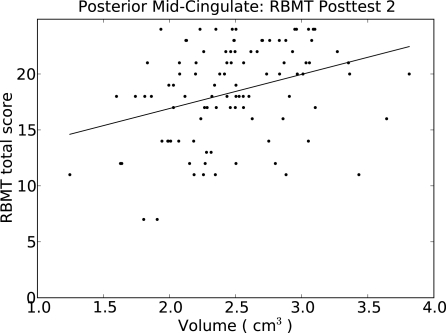
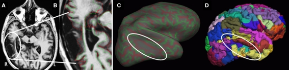
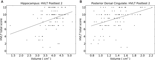
Cognitive deficits following traumatic brain injury (TBI) commonly include difficulties with memory, attention, and executive dysfunction. These deficits are amenable to cognitive rehabilitation, but optimally selecting rehabilitation programs for individual patients remains a challenge. Recent methods for quantifying regional brain morphometry allow for automated quantification of tissue volumes in numerous distinct brain structures. We hypothesized that such quantitative structural information could help identify individuals more or less likely to benefit from memory rehabilitation. Fifty individuals with TBI of all severities who reported having memory difficulties first underwent structural MRI scanning. They then participated in a 12 session memory rehabilitation program emphasizing internal memory strategies (I-MEMS). Primary outcome measures (HVLT, RBMT) were collected at the time of the MRI scan, immediately following therapy, and again at 1-month post-therapy. Regional brain volumes were used to predict outcome, adjusting for standard predictors (e.g., injury severity, age, education, pretest scores). We identified several brain regions that provided significant predictions of rehabilitation outcome, including the volume of the hippocampus, the lateral prefrontal cortex, the thalamus, and several subregions of the cingulate cortex. The prediction range of regional brain volumes were in some cases nearly equal in magnitude to prediction ranges provided by pretest scores on the outcome variable. We conclude that specific cerebral networks including these regions may contribute to learning during I-MEMS rehabilitation, and suggest that morphometric measures may provide substantial predictive value for rehabilitation outcome in other cognitive interventions as well.
创伤性脑损伤(TBI)后的认知缺陷通常包括记忆、注意力和执行功能障碍方面的困难。这些缺陷适合进行认知康复,但为个体患者优化选择康复方案仍然是一项挑战。最近用于量化局部脑形态测量的方法能够自动量化众多不同脑结构中的组织体积。我们假设这种定量结构信息有助于识别更有可能或不太可能从记忆康复中受益的个体。五十名报告有记忆困难的各种严重程度的TBI患者首先接受了结构MRI扫描。然后他们参加了一个为期12节的记忆康复计划,该计划强调内部记忆策略(I-MEMS)。主要结局指标(HVLT、RBMT)在MRI扫描时、治疗后立即以及治疗后1个月再次收集。使用局部脑体积来预测结局,并对标准预测因素(如损伤严重程度、年龄、教育程度、预测试分数)进行调整。我们确定了几个对康复结局有显著预测作用的脑区,包括海马体、外侧前额叶皮质、丘脑以及扣带回皮质的几个亚区。局部脑体积的预测范围在某些情况下几乎与结局变量的预测试分数提供的预测范围大小相当。我们得出结论,包括这些区域在内的特定脑网络可能有助于I-MEMS康复期间的学习,并表明形态测量指标可能对其他认知干预中的康复结局也具有重要的预测价值。