Ayaki Masahiko, Iwasawa Atsuo, Inoue Yoichi
Department of Ophthalmology, Saitama National Hospital, Wako, Japan.
Clin Ophthalmol. 2010 Oct 21;4:1217-22. doi: 10.2147/OPTH.S13708.
The toxicity of antiglaucoma medications to ocular surface cells has been evaluated extensively; however, the toxicity to corneal endothelial cells (CECs) remains elusive. Our aim is to evaluate the toxicity of antiglaucoma medications to CECs using an in vitro toxicity assay.
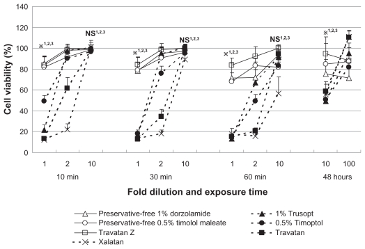
Primary cultures of human (H) CECs derived from eye bank specimens were established. Following exposure of HCECs to test solutions for 10, 30, or 60 minutes, or 48 hours, we measured cell viability using a WST-1 assay. Test solutions were diluted in culture media and included 0.5% Timoptol(®), preservative-free 0.5% timolol maleate, 1% Trusopt(®), preservative-free 1% dorzolamide, Travatan(®), Travatan Z(®), Xalatan(®), and benzalkonium chloride (BAK). To assess cell viability, the value of the test culture well after treatment was expressed as a percentage of that of the control well. Toxicity of each solution was compared using the cell viability score (CVS).
After exposure to 10-fold dilutions of test solutions for 48 hours, HCEC viabilities were 48.5% for 0.5% Timoptol, 80.9% for preservative-free 0.5% timolol maleate, 47.0% for 1% Trusopt, 71.7% for preservative-free 1% dorzolamide, 55.5% for Travatan, 88.5% for Travatan Z, and 52.5% for Xalatan. Exposure to test solutions diluted 100-fold or more resulted in HCEC viabilities >80%, with the exception of preservative-free 1% dorzolamide, which resulted in a viability of 72.0% at a dilution of 100-fold. Based on CVS, the order of cell viability was Travatan Z ≥ preservative-free timolol maleate = preservative-free dorzolamide > 0.5% Timoptol = 1% Trusopt > Travatan ≥ Xalatan. Assessment of the combined effect of drug and BAK revealed that latanoprost reduced the toxicity of BAK.
Antiglaucoma eye drops produced HCEC toxicity that appeared to depend on the presence of BAK. Because dilution of the antiglaucoma solutions resulted in markedly lower HCEC toxicity, HCEC damage due to antiglaucoma medication may occur only in rare cases. The CVS was useful for comparison of the toxicity of the drugs.
抗青光眼药物对眼表细胞的毒性已得到广泛评估;然而,其对角膜内皮细胞(CECs)的毒性仍不清楚。我们的目的是使用体外毒性试验评估抗青光眼药物对CECs的毒性。
建立了源自眼库标本的人(H)CECs原代培养物。将HCECs暴露于测试溶液10、30或60分钟或48小时后,我们使用WST-1试验测量细胞活力。测试溶液在培养基中稀释,包括0.5%噻吗洛尔(Timoptol®)、无防腐剂的0.5%马来酸噻吗洛尔、1%布林佐胺(Trusopt®)、无防腐剂的1%多佐胺、曲伏前列素(Travatan®)、曲伏前列素Z(Travatan Z®)、拉坦前列素(Xalatan®)和苯扎氯铵(BAK)。为评估细胞活力,处理后测试培养孔的值表示为对照孔值的百分比。使用细胞活力评分(CVS)比较每种溶液的毒性。
在将测试溶液稀释10倍后暴露48小时后,0.5%噻吗洛尔组HCEC活力为48.5%,无防腐剂的0.5%马来酸噻吗洛尔组为80.9%,1%布林佐胺组为47.0%,无防腐剂的1%多佐胺组为71.7%,曲伏前列素组为55.5%,曲伏前列素Z组为88.5%,拉坦前列素组为52.5%。暴露于稀释100倍或更高倍数的测试溶液导致HCEC活力>80%,无防腐剂的1%多佐胺除外,其在100倍稀释时活力为72.0%。基于CVS,细胞活力顺序为曲伏前列素Z≥无防腐剂的马来酸噻吗洛尔=无防腐剂的多佐胺>0.5%噻吗洛尔=1%布林佐胺>曲伏前列素≥拉坦前列素。对药物和BAK联合作用的评估显示,拉坦前列素降低了BAK的毒性。
抗青光眼滴眼液产生的HCEC毒性似乎取决于BAK的存在。由于抗青光眼溶液的稀释导致HCEC毒性显著降低,抗青光眼药物引起的HCEC损伤可能仅在极少数情况下发生。CVS有助于比较药物的毒性。