Image Sciences Institute, University Medical Centre Utrecht, Utrecht, The Netherlands.
Eur J Nucl Med Mol Imaging. 2011 Mar;38(3):552-61. doi: 10.1007/s00259-010-1637-4. Epub 2010 Nov 10.
Small-animal single photon emission computed tomography (SPECT) with focused multi-pinhole collimation geometries allows scanning modes in which large amounts of photons can be collected from specific volumes of interest. Here we present new tools that improve targeted imaging of specific organs and tumours, and validate the effects of improved targeting of the pinhole focus.
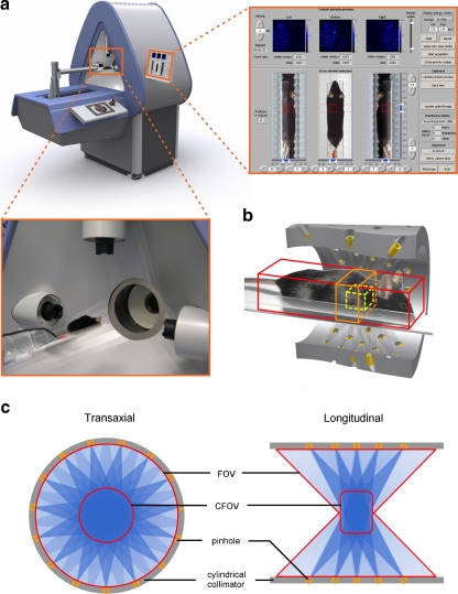
A SPECT system with 75 pinholes and stationary detectors was used (U-SPECT-II). An XYZ stage automatically translates the animal bed with a specific sequence in order to scan a selected volume of interest. Prior to stepping the animal through the collimator, integrated webcams acquire images of the animal. Using sliders, the user designates the desired volume to be scanned (e.g. a xenograft or specific organ) on these optical images. Optionally projections of an atlas are overlaid semiautomatically to locate specific organs. In order to assess the effects of more targeted imaging, scans of a resolution phantom and a mouse myocardial phantom, as well as in vivo mouse cardiac and tumour scans, were acquired with increased levels of targeting. Differences were evaluated in terms of count yield, hot rod visibility and contrast-to-noise ratio.
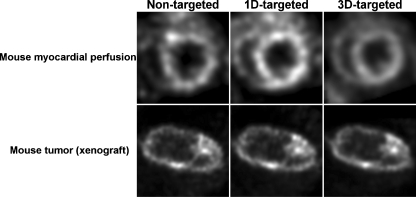
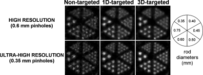
By restricting focused SPECT scans to a 1.13-ml resolution phantom, count yield was increased by a factor 3.6, and visibility of small structures was significantly enhanced. At equal noise levels, the small-lesion contrast measured in the myocardial phantom was increased by 42%. Noise in in vivo images of a tumour and the mouse heart was significantly reduced.
Targeted pinhole SPECT improves images and can be used to shorten scan times. Scan planning with optical cameras provides an effective tool to exploit this principle without the necessity for additional X-ray CT imaging.
采用聚焦多针孔准直几何结构的小动物单光子发射计算机断层扫描(SPECT)可以从特定感兴趣体积中采集大量光子。在此,我们介绍了新的工具,可改善对特定器官和肿瘤的靶向成像,并验证了改善针孔焦点靶向的效果。
使用配备 75 个针孔和固定探测器的 SPECT 系统(U-SPECT-II)。XYZ 载物台可自动以特定顺序平移动物床,以扫描选定的感兴趣体积。在将动物穿过准直器之前,集成的网络摄像头会获取动物的图像。使用滑块,用户在这些光学图像上指定要扫描的目标体积(例如,异种移植物或特定器官)。可选地,图谱的投影会半自动叠加以定位特定器官。为了评估更具针对性的成像效果,我们增加了靶向水平,对分辨率体模和小鼠心肌体模以及体内小鼠心脏和肿瘤扫描进行了扫描。通过限制聚焦 SPECT 扫描到 1.13ml 的分辨率体模,计数产量增加了 3.6 倍,并且可以显著增强小结构的可见度。在相同噪声水平下,心肌体模中小病变的对比度增加了 42%。肿瘤和小鼠心脏的体内图像噪声显著降低。
靶向针孔 SPECT 可以改善图像质量,并可用于缩短扫描时间。使用光学相机进行扫描规划是一种有效的工具,可以利用该原理,而无需额外的 X 射线 CT 成像。