Kojima Takashi, Matsumoto Yukihiro, Dogru Murat, Tsubota Kazuo
Johnson & Johnson Department of Ocular Surface and Visual Optics, Keio University School of Medicine, Tokyo, Japan.
Mol Vis. 2010 Nov 19;16:2457-64.
To evaluate the applicability of in vivo laser scanning confocal microscopy as a tool of conjunctival cytology in a prospective case-control study.
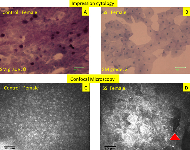
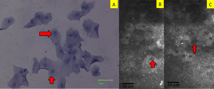
Nineteen right eyes of 19 Sjogren's syndrome dry eye patients (19 females; mean age: 55.8±15 years), and 18 right eyes of 18 normal healthy control subjects (12 females and 6 males; mean age: 50.8±14 years) were evaluated in this study. The eyes were analyzed by the Heidelberg retina tomography (HRTII)/Rostock cornea module (RCM). Ocular surface and tear function tests including vital stainings (fluorescein and Rose Bengal), Schirmer test, tear film break up time (BUT), and conjunctival impression cytology were performed. After obtaining the confocal microscopy images, the mean individual epithelial cell area (MIECA), and nucleocytoplasmic (N/C) ratio were analyzed. The correlation between confocal microscopy and impression cytology parameters was also investigated.
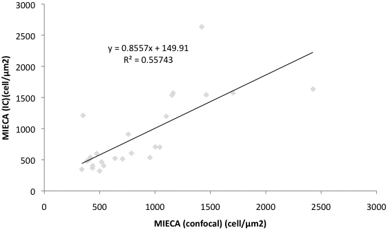
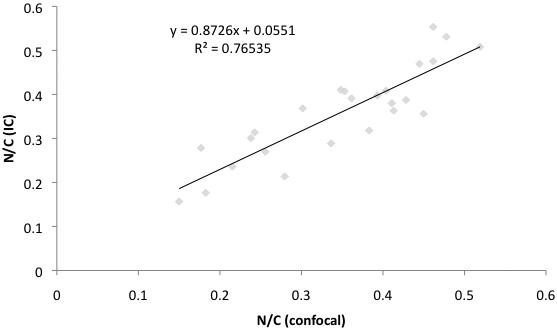
The BUT, Schirmer test values, vital staining scores and squamous metaplasia grades in impression cytology were significantly worse in dry eye patients compared to controls (p<0.0001). The MIECA and the mean N/C ratios were worse in dry eye subjects compared to controls both in impression cytology and in vivo confocal microscopy (p<0.0001) with no significant differences between these parameters when the two examination techniques were compared. The MIECA and N/C ratio in conjunctival impression cytology showed significant correlation with the corresponding confocal microscopy parameters (MIECA, r2:0.557 ; N/C, r2:0.765).
Laser scanning confocal microscopy seems to be an efficient non-invasive tool in the evaluation of phenotypic alterations of the conjunctival epithelium in dry eye disease. N/C ratio and MIECA appear to be two promising and new parameters of in vivo confocal cytology in the assessment of the ocular surface in dry eye disease.
在一项前瞻性病例对照研究中评估体内激光扫描共聚焦显微镜作为结膜细胞学检查工具的适用性。
本研究评估了19例干燥综合征干眼患者(19名女性;平均年龄:55.8±15岁)的19只右眼,以及18名正常健康对照者(12名女性和6名男性;平均年龄:50.8±14岁)的18只右眼。使用海德堡视网膜断层扫描(HRTII)/罗斯托克角膜模块(RCM)对眼睛进行分析。进行了眼表和泪液功能测试,包括活体染色(荧光素和孟加拉玫瑰红)、泪液分泌试验、泪膜破裂时间(BUT)和结膜印片细胞学检查。获取共聚焦显微镜图像后,分析平均单个上皮细胞面积(MIECA)和核质比(N/C)。还研究了共聚焦显微镜与印片细胞学参数之间的相关性。
与对照组相比,干眼患者的BUT、泪液分泌试验值、活体染色评分和印片细胞学中的鳞状化生分级明显更差(p<0.0001)。在印片细胞学和体内共聚焦显微镜检查中,干眼受试者的MIECA和平均N/C比均比对照组更差(p<0.0001),当比较这两种检查技术时,这些参数之间无显著差异。结膜印片细胞学中的MIECA和N/C比与相应的共聚焦显微镜参数显示出显著相关性(MIECA,r2:0.557;N/C,r2:0.765)。
激光扫描共聚焦显微镜似乎是评估干眼疾病中结膜上皮表型改变的一种有效的非侵入性工具。N/C比和MIECA似乎是体内共聚焦细胞学在评估干眼疾病眼表时两个有前景的新参数。