• 文献检索
  • 文档翻译
  • 深度研究
  • 学术资讯
  • Suppr Zotero 插件Zotero 插件
  • 邀请有礼
  • 套餐&价格
  • 历史记录
应用&插件
Suppr Zotero 插件Zotero 插件浏览器插件Mac 客户端Windows 客户端微信小程序
定价
高级版会员购买积分包购买API积分包
服务
文献检索文档翻译深度研究API 文档MCP 服务
关于我们
关于 Suppr公司介绍联系我们用户协议隐私条款
关注我们

Suppr 超能文献

核心技术专利:CN118964589B侵权必究
粤ICP备2023148730 号-1Suppr @ 2026

文献检索

告别复杂PubMed语法,用中文像聊天一样搜索,搜遍4000万医学文献。AI智能推荐,让科研检索更轻松。

立即免费搜索

文件翻译

保留排版,准确专业,支持PDF/Word/PPT等文件格式,支持 12+语言互译。

免费翻译文档

深度研究

AI帮你快速写综述,25分钟生成高质量综述,智能提取关键信息,辅助科研写作。

立即免费体验

胸膜良性孤立性纤维瘤的影像学表现:图文综述

Imaging of benign solitary fibrous tumor of the pleura: a pictorial essay.

作者信息

Cardinale Luciano, Ardissone Francesco, Garetto Irene, Marci Valerio, Volpicelli Giovanni, Solitro Federica, Fava Cesare

机构信息

Institute of Radiology, University of Turin;

出版信息

Rare Tumors. 2010 Mar 31;2(1):e1. doi: 10.4081/rt.2010.e1.

DOI:10.4081/rt.2010.e1
PMID:21139938
原文链接:https://pmc.ncbi.nlm.nih.gov/articles/PMC2994496/
Abstract

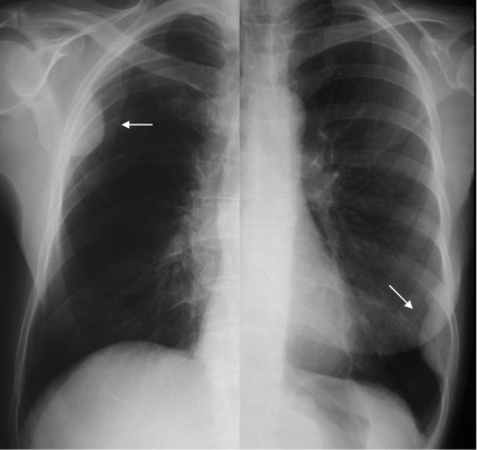
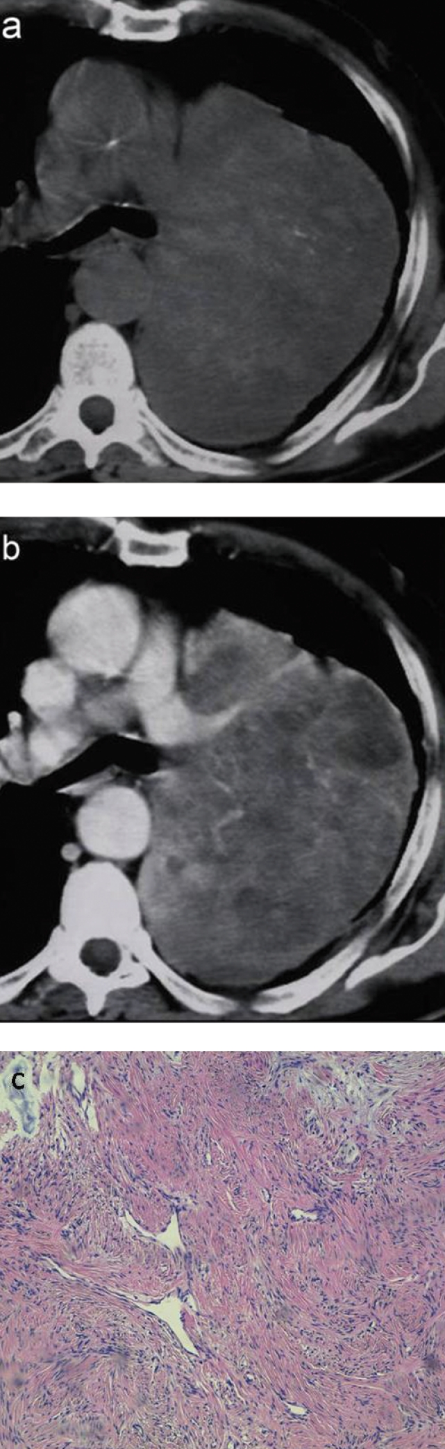
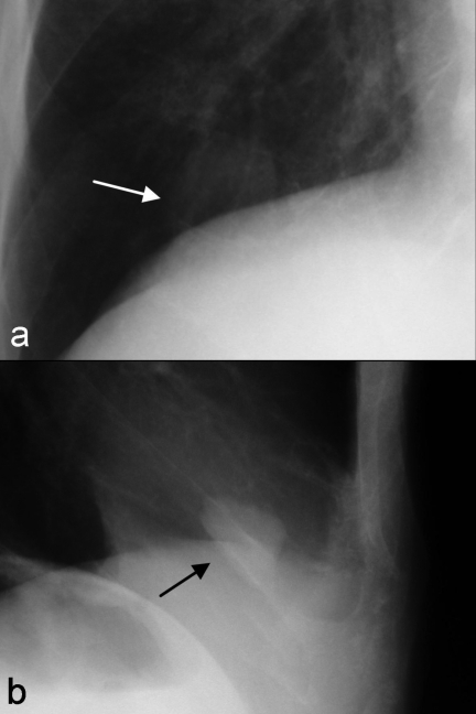
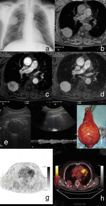
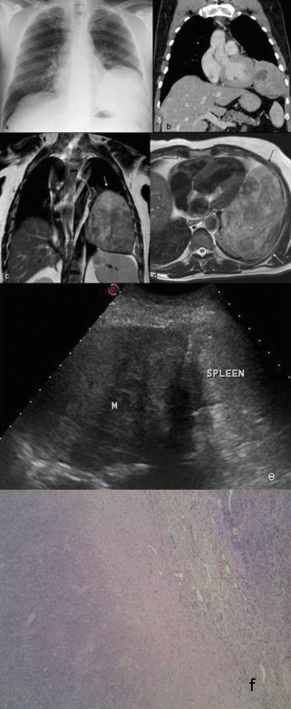
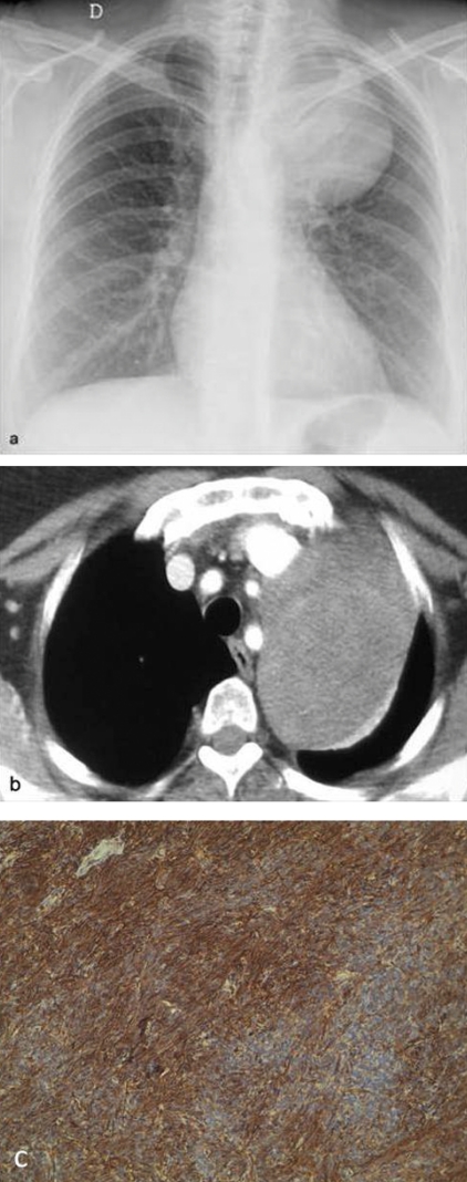
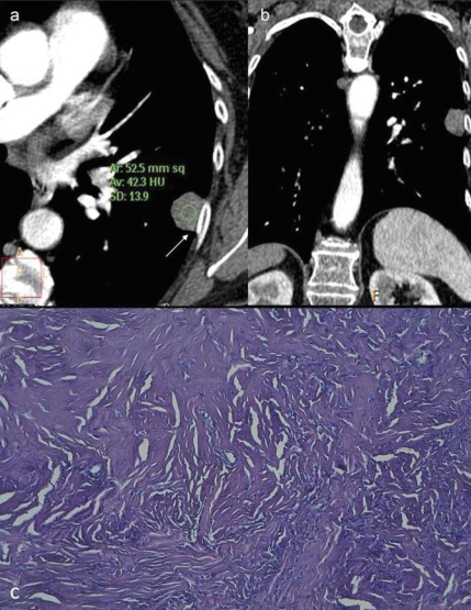
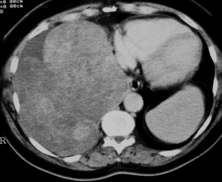
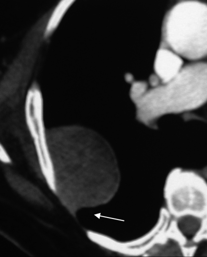
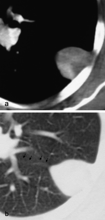
Solitary fibrous tumor of the pleura (SFTP) is a mesenchymal tumor that tends to involve the pleura, and is also described in other thoracic and extrathoracic sites. SFTP usually presents as a peripheral mass abutting the pleural surface, to which it is attached by a broad base or by a pedicle that allows it to be mobile. SFTPs exist in benign and malignant forms. A precise pre-operative diagnosis can be arrived at with a cutting-needle biopsy, although most cases are diagnosed with postoperative histology and immunohistochemical analysis. In this pictorial essay, we review a large series of cases, with emphasis on the radiographic appearance of these lesions and their findings from computed tomography, magnetic resonance imaging, ultrasonography and positron emission tomography.

摘要

胸膜孤立性纤维瘤(SFTP)是一种间叶性肿瘤,倾向于累及胸膜,也可见于其他胸部和胸外部位。SFTP通常表现为紧贴胸膜表面的周围型肿块,通过宽基底或蒂与胸膜相连,使其具有可移动性。SFTP有良性和恶性两种形式。虽然大多数病例是通过术后组织学和免疫组化分析确诊的,但通过切割针活检可作出精确的术前诊断。在这篇影像专题文章中,我们回顾了大量病例,重点介绍这些病变的影像学表现以及计算机断层扫描、磁共振成像、超声检查和正电子发射断层扫描的结果。

https://cdn.ncbi.nlm.nih.gov/pmc/blobs/c219/2994496/e80eb70dceff/rt-2010-1-e1-g011.jpg
https://cdn.ncbi.nlm.nih.gov/pmc/blobs/c219/2994496/9ca4c9b35ed6/rt-2010-1-e1-g001.jpg
https://cdn.ncbi.nlm.nih.gov/pmc/blobs/c219/2994496/73c0adf0c651/rt-2010-1-e1-g002.jpg
https://cdn.ncbi.nlm.nih.gov/pmc/blobs/c219/2994496/81fb9f9bab5e/rt-2010-1-e1-g003.jpg
https://cdn.ncbi.nlm.nih.gov/pmc/blobs/c219/2994496/1e4e99f732f4/rt-2010-1-e1-g004.jpg
https://cdn.ncbi.nlm.nih.gov/pmc/blobs/c219/2994496/981a00800a4e/rt-2010-1-e1-g005.jpg
https://cdn.ncbi.nlm.nih.gov/pmc/blobs/c219/2994496/3175ca5151d2/rt-2010-1-e1-g006.jpg
https://cdn.ncbi.nlm.nih.gov/pmc/blobs/c219/2994496/ca69ec2a827d/rt-2010-1-e1-g007.jpg
https://cdn.ncbi.nlm.nih.gov/pmc/blobs/c219/2994496/21aa31828f1f/rt-2010-1-e1-g008.jpg
https://cdn.ncbi.nlm.nih.gov/pmc/blobs/c219/2994496/fdfe1ca917f5/rt-2010-1-e1-g009.jpg
https://cdn.ncbi.nlm.nih.gov/pmc/blobs/c219/2994496/406f6f797c27/rt-2010-1-e1-g010.jpg
https://cdn.ncbi.nlm.nih.gov/pmc/blobs/c219/2994496/e80eb70dceff/rt-2010-1-e1-g011.jpg

相似文献

1
Imaging of benign solitary fibrous tumor of the pleura: a pictorial essay.胸膜良性孤立性纤维瘤的影像学表现:图文综述
Rare Tumors. 2010 Mar 31;2(1):e1. doi: 10.4081/rt.2010.e1.
2
CT signs, patterns and differential diagnosis of solitary fibrous tumors of the pleura.胸膜孤立性纤维瘤的 CT 征象、类型及鉴别诊断。
J Thorac Dis. 2010 Mar;2(1):21-5.
3
Fibrous tumour of the pleura (SFTP): a proteiform disease. Clinical, histological and atypical radiological patterns selected among our cases.胸膜纤维瘤(孤立性纤维性肿瘤):一种形态多样的疾病。从我们的病例中选取的临床、组织学及非典型放射学表现
Radiol Med. 2009 Mar;114(2):204-15. doi: 10.1007/s11547-008-0345-7. Epub 2008 Dec 11.
4
Resection of giant malignant solitary fibrous pleural tumor after interventional embolization: a case report and literature review.介入栓塞后切除巨大恶性孤立性纤维性胸膜肿瘤:病例报告及文献复习。
J Cardiothorac Surg. 2022 May 31;17(1):134. doi: 10.1186/s13019-022-01881-z.
5
Solitary Fibrous Tumor of the Pleura: A Rare Cause of Pleural Mass.胸膜孤立性纤维瘤:胸膜肿块的罕见病因。
Am J Case Rep. 2015 Dec 3;16:854-7. doi: 10.12659/ajcr.895289.
6
The utility of 18F-FDG PET/CT in solitary fibrous tumors of the pleura.18F-FDG PET/CT在胸膜孤立性纤维瘤中的应用价值。
Rev Esp Med Nucl Imagen Mol. 2016 May-Jun;35(3):165-70. doi: 10.1016/j.remn.2015.10.003. Epub 2015 Nov 27.
7
Giant benign solitary fibrous tumor of the pleura: PET/CT findings.胸膜巨大良性孤立性纤维瘤:PET/CT 表现。
Clin Nucl Med. 2012 Jul;37(7):702-4. doi: 10.1097/RLU.0b013e31824c6054.
8
Solitary fibrous tumour of pleura: CT differentiation of benign and malignant types.胸膜孤立性纤维瘤:良恶性类型的CT鉴别
Clin Radiol. 2017 Sep;72(9):796.e9-796.e17. doi: 10.1016/j.crad.2017.03.028. Epub 2017 May 8.
9
Radiology-Pathology Conference: 18F FDG PET-CT imaging of solitary fibrous tumor of the pleura.放射科-病理科联合会诊:18F-FDG PET-CT 成像在胸膜孤立性纤维瘤中的应用。
Clin Imaging. 2013 May-Jun;37(3):598-601. doi: 10.1016/j.clinimag.2012.09.020. Epub 2012 Oct 30.
10
Multiple solitary fibrous tumors of the pleura with multicentric and unilateral involvement: a case report.多中心单侧累及的胸膜多发性孤立性纤维瘤:病例报告
Surg Case Rep. 2023 Jul 26;9(1):134. doi: 10.1186/s40792-023-01717-5.

引用本文的文献

1
Surgery for Solitary Fibrous Tumors of the Pleura: A Review of the Available Evidence.胸膜孤立性纤维瘤的手术治疗:现有证据综述
Cancers (Basel). 2023 Aug 18;15(16):4166. doi: 10.3390/cancers15164166.
2
A Case of a Large and Rare Incidental Pleural Tumor in an Elderly Female.一位老年女性中发现的巨大罕见偶然性胸膜肿瘤病例。
Cureus. 2023 Jul 20;15(7):e42198. doi: 10.7759/cureus.42198. eCollection 2023 Jul.
3
Imaging Features of Various Benign and Malignant Tumors and Tumorlike Conditions of the Pleura: A Pictorial Review.胸膜各种良性和恶性肿瘤及肿瘤样病变的影像学特征:图谱综述

本文引用的文献

1
Recurrent localized fibrous tumor of the pleura.复发性局限性胸膜纤维瘤
Ann Thorac Surg. 2006 Jul;82(1):342-5. doi: 10.1016/j.athoracsur.2005.09.057.
2
CT features of solitary fibrous tumour of the pleura: experience in 26 patients.胸膜孤立性纤维瘤的CT特征:26例患者的经验
Radiol Med. 2006 Aug;111(5):640-50. doi: 10.1007/s11547-006-0062-z. Epub 2006 Jun 29.
3
MR imaging of solitary fibrous tumors in the head and neck.头颈部孤立性纤维瘤的磁共振成像
Taehan Yongsang Uihakhoe Chi. 2020 Sep;81(5):1109-1120. doi: 10.3348/jksr.2019.0165. Epub 2020 Aug 13.
4
Pleural nodule with osteal protrusion anterior to the rib tubercle: a case report.肋骨结节前方伴有骨质突出的胸膜结节:一例报告
Radiol Case Rep. 2021 Jun 8;16(8):2091-2094. doi: 10.1016/j.radcr.2021.05.026. eCollection 2021 Aug.
5
Solitary fibrous tumor of the pleura: a rare cause of elevation of the right lung base.胸膜孤立性纤维瘤:右肺底抬高的罕见原因。
J Bras Pneumol. 2019 Feb 28;45(1):e20180006. doi: 10.1590/1806-3713/e20180006.
6
A case report of solitary fibrous tumor of the pleura: ultrasound diagnostic features.胸膜孤立性纤维瘤病例报告:超声诊断特征
J Med Ultrason (2001). 2012 Apr;39(2):97-100. doi: 10.1007/s10396-011-0338-3. Epub 2012 Jan 6.
7
How I do it: lung ultrasound.我的做法:肺部超声检查。
Cardiovasc Ultrasound. 2014 Jul 4;12:25. doi: 10.1186/1476-7120-12-25.
8
Solitary fibrous tumour of the pleura: a moving target.胸膜孤立性纤维瘤:一个不断变化的目标。
Can Respir J. 2014 May-Jun;21(3):145. doi: 10.1155/2014/979053. Epub 2014 Apr 7.
9
Solitary fibrous tumor of the pleura.胸膜孤立性纤维瘤。
Med Oncol. 2013;30(2):573. doi: 10.1007/s12032-013-0573-0. Epub 2013 Apr 11.
10
CT Imaging of Solitary fibrous tumour of the pleura (SFTP): Typical Patterns and pitfalls.胸膜孤立性纤维瘤(SFTP)的CT成像:典型表现及陷阱
J Thorac Dis. 2012 Apr 1;4(2):112-3. doi: 10.3978/j.issn.2072-1439.2011.09.01.
Korean J Radiol. 2005 Jul-Sep;6(3):136-42. doi: 10.3348/kjr.2005.6.3.136.
4
[(18)F]-FDG PET and localized fibrous mesothelioma.
Lung. 2003;181(1):49-54. doi: 10.1007/s00408-003-1002-5.
5
From the archives of the AFIP: Localized fibrous tumor of the pleura.源自武装部队病理研究所档案:胸膜局限性纤维瘤。
Radiographics. 2003 May-Jun;23(3):759-83. doi: 10.1148/rg.233025165.
6
Solitary fibrous tumors of the pleura.胸膜孤立性纤维瘤
Ann Thorac Surg. 2002 Jul;74(1):285-93. doi: 10.1016/s0003-4975(01)03374-4.
7
Solitary fibrous tumor of the pleura: MR appearance and enhancement pattern.胸膜孤立性纤维瘤:磁共振成像表现及强化方式
J Comput Assist Tomogr. 2002 Mar-Apr;26(2):174-9. doi: 10.1097/00004728-200203000-00002.
8
Solitary fibrous tumor: a study of cytologic features of six cases diagnosed by fine-needle aspiration.
Diagn Cytopathol. 2001 Sep;25(3):172-6. doi: 10.1002/dc.2032.
9
Localized benign fibrous tumors of the pleura.
AJR Am J Roentgenol. 1997 Sep;169(3):683-6. doi: 10.2214/ajr.169.3.9275877.
10
Localized benign fibrous tumors of the pleura: MR appearance.胸膜局限性良性纤维瘤:磁共振成像表现
J Comput Assist Tomogr. 1997 Jan-Feb;21(1):115-20. doi: 10.1097/00004728-199701000-00023.