• 文献检索
  • 文档翻译
  • 深度研究
  • 学术资讯
  • Suppr Zotero 插件Zotero 插件
  • 邀请有礼
  • 套餐&价格
  • 历史记录
应用&插件
Suppr Zotero 插件Zotero 插件浏览器插件Mac 客户端Windows 客户端微信小程序
定价
高级版会员购买积分包购买API积分包
服务
文献检索文档翻译深度研究API 文档MCP 服务
关于我们
关于 Suppr公司介绍联系我们用户协议隐私条款
关注我们

Suppr 超能文献

核心技术专利:CN118964589B侵权必究
粤ICP备2023148730 号-1Suppr @ 2026

文献检索

告别复杂PubMed语法,用中文像聊天一样搜索,搜遍4000万医学文献。AI智能推荐,让科研检索更轻松。

立即免费搜索

文件翻译

保留排版,准确专业,支持PDF/Word/PPT等文件格式,支持 12+语言互译。

免费翻译文档

深度研究

AI帮你快速写综述,25分钟生成高质量综述,智能提取关键信息,辅助科研写作。

立即免费体验

小动物在 7T 下的脂肪分割:初步研究。

Adipose segmentation in small animals at 7T: a preliminary study.

机构信息

Department of Radiology, University of Southern California, Childrens Hospital Los Angeles, Los Angeles, USA.

出版信息

BMC Genomics. 2010 Dec 1;11 Suppl 3(Suppl 3):S9. doi: 10.1186/1471-2164-11-S3-S9.

DOI:10.1186/1471-2164-11-S3-S9
PMID:21143791
原文链接:https://pmc.ncbi.nlm.nih.gov/articles/PMC2999354/
Abstract

BACKGROUND

Small animal MRI at 7 Tesla (T) provides a useful tool for adiposity research. For adiposity researchers, separation of fat from surrounding tissues and its subsequent quantitative or semi- quantitative analysis is a key task. This is a relatively new field and a priori it cannot be known which specific biological questions related to fat deposition will be relevant in a specific study. Thus it is impossible to predict what accuracy and what spatial resolution will be required in all cases and even difficult what accuracy and resolution will be useful in most cases. However the pragmatic time constraints and the practical resolution ranges are known for small animal imaging at 7T. Thus we have used known practical constraints to develop a method for fat volume analysis based on an optimized image acquisition and image post processing pair.

METHODS

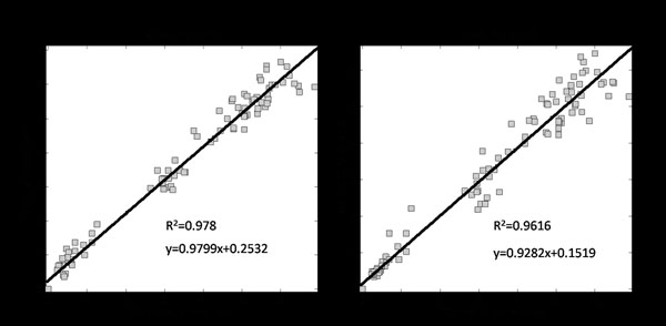
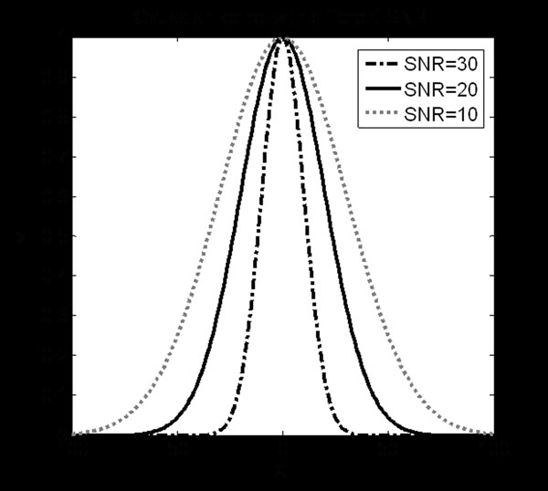
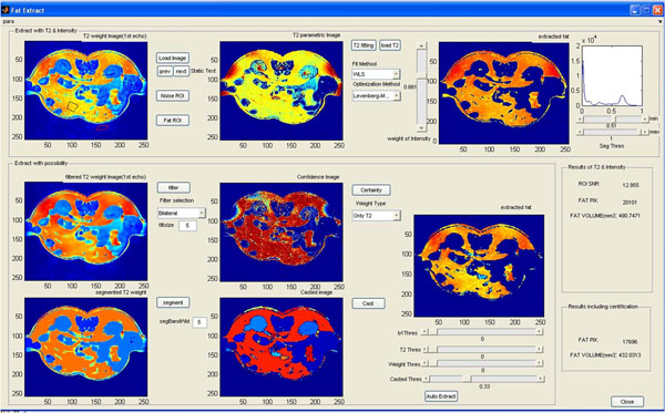
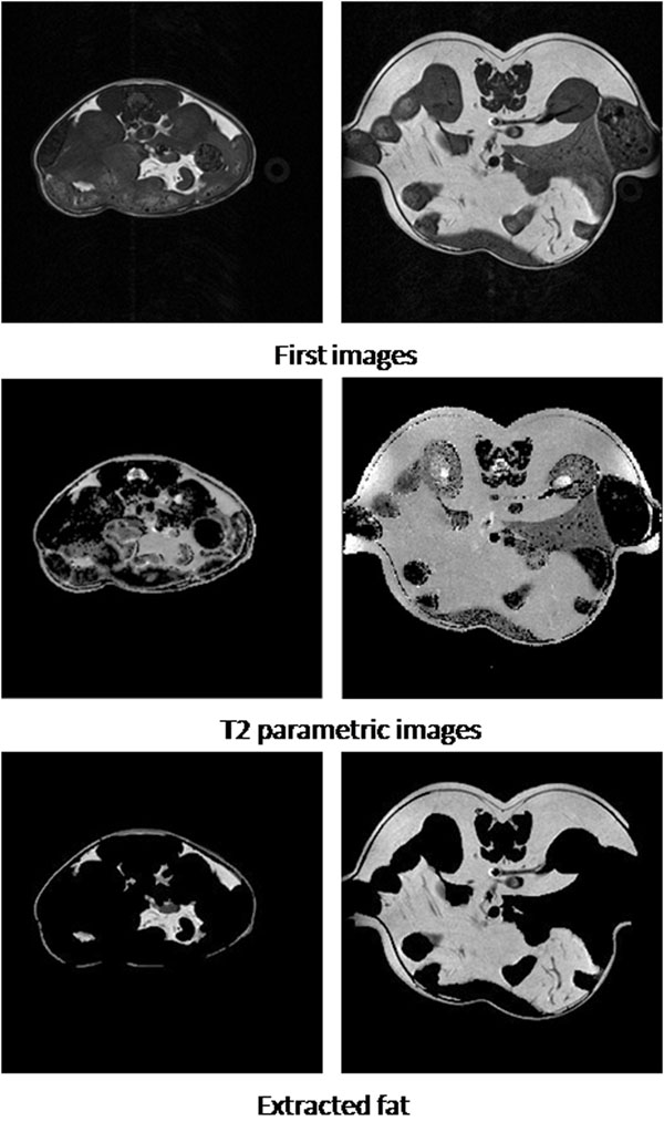
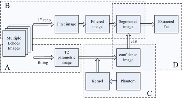
We designed a fat segmentation method based on optimizing a variety of factors relevant to small animal imaging at 7T. In contrast to most previously described MRI methods based on signal intensity of T1 weighted image alone, we chose to use parametric images based on Multi-spin multi-echo (MSME) Bruker pulse sequence which has proven to be particularly robust in our laboratory over the last several years. The sequence was optimized on a T1 basis to emphasize the signal. T2 relaxation times can be calculated from the multi echo data and we have done so on a pixel by pixel basis for the initial step in the post processing methodology. The post processing consists of parallel paths. On one hand, the weighted image is precisely divided into different regions with optimized smoothing and segmentation methods; and on the other hand, a confidence image is deduced from the parametric image according to the distribution of relaxation time relationship of typical adipose. With the assistance of the confidence image, a useful software feature was implemented to which enhances the data and in the end results in a more reliable and flexible method for adipose evaluation.

RESULTS

In this paper, we describe how we arrived at our recommended procedures and key aspects of the post-processing steps. The feasibility of the proposed method is tested on both simulated and real data in this preliminary research. A research tool was created to help researchers segment out fat even when the anatomical information is of low quality making it difficult to distinguish between fat and non-fat. In addition, tool is designed to allow the operator to make adjustments to many of the key steps for comparison purposes and to quantitatively assess the difference these changes make. Ultimately our flexible software lets the researcher define key aspects of the fat segmentation and quantification.

CONCLUSIONS

Combining the full T2 parametric information with the optimized first echo image information, the research tool enhances the reliability of the results while providing more flexible operations than previous methods. The innovation in the method is to pair an optimized and very specific image acquisition technique to a flexible but tuned image post processing method. The separation of the fat is aided by the confidence distribution of regions produced on a scale relevant to and dictated by practical aspects of MRI at 7T.

https://cdn.ncbi.nlm.nih.gov/pmc/blobs/31ff/2999354/b18fe0399d9c/1471-2164-11-S3-S9-10.jpg
https://cdn.ncbi.nlm.nih.gov/pmc/blobs/31ff/2999354/ec5680ba10be/1471-2164-11-S3-S9-1.jpg
https://cdn.ncbi.nlm.nih.gov/pmc/blobs/31ff/2999354/8a1f408d2219/1471-2164-11-S3-S9-2.jpg
https://cdn.ncbi.nlm.nih.gov/pmc/blobs/31ff/2999354/ee2ec9553693/1471-2164-11-S3-S9-3.jpg
https://cdn.ncbi.nlm.nih.gov/pmc/blobs/31ff/2999354/ae7ba8ad1dfc/1471-2164-11-S3-S9-4.jpg
https://cdn.ncbi.nlm.nih.gov/pmc/blobs/31ff/2999354/e83f997c9c1b/1471-2164-11-S3-S9-5.jpg
https://cdn.ncbi.nlm.nih.gov/pmc/blobs/31ff/2999354/065b5e0c7456/1471-2164-11-S3-S9-6.jpg
https://cdn.ncbi.nlm.nih.gov/pmc/blobs/31ff/2999354/16d87135d6fb/1471-2164-11-S3-S9-7.jpg
https://cdn.ncbi.nlm.nih.gov/pmc/blobs/31ff/2999354/38c881c1fac4/1471-2164-11-S3-S9-8.jpg
https://cdn.ncbi.nlm.nih.gov/pmc/blobs/31ff/2999354/6d0e778e7956/1471-2164-11-S3-S9-9.jpg
https://cdn.ncbi.nlm.nih.gov/pmc/blobs/31ff/2999354/b18fe0399d9c/1471-2164-11-S3-S9-10.jpg
摘要

背景

在 7 特斯拉(T)下进行小动物 MRI 提供了用于肥胖症研究的有用工具。对于肥胖症研究人员来说,将脂肪与周围组织分离并对其进行定量或半定量分析是一项关键任务。这是一个相对较新的领域,不能事先知道与脂肪沉积相关的特定生物学问题将在特定研究中相关。因此,无法预测在所有情况下都需要什么样的准确性和空间分辨率,甚至难以预测在大多数情况下什么样的准确性和分辨率将是有用的。然而,在 7T 小动物成像中已知实际的时间限制和实际的分辨率范围。因此,我们已经利用已知的实际限制,基于优化的图像采集和图像后处理对,开发了一种脂肪体积分析方法。

方法

我们设计了一种基于优化与 7T 小动物成像相关的各种因素的脂肪分割方法。与大多数以前基于单独 T1 加权图像信号强度描述的 MRI 方法不同,我们选择使用基于多自旋多回波(MSME)Bruker 脉冲序列的参数图像,该方法在过去几年中在我们的实验室中已被证明特别可靠。该序列基于 T1 进行了优化以强调信号。可以从多回波数据中计算出 T2 弛豫时间,并且我们已经在像素的基础上进行了计算,作为后处理方法学的初始步骤。后处理包括平行路径。一方面,使用优化的平滑和分割方法精确地将加权图像划分为不同的区域;另一方面,根据典型脂肪的弛豫时间关系分布,从参数图像中推断出置信度图像。借助置信度图像,实现了一个有用的软件功能,该功能增强了数据,并最终为脂肪评估提供了更可靠和灵活的方法。

结果

在本文中,我们描述了如何得出我们推荐的程序以及后处理步骤的关键方面。在这项初步研究中,使用模拟和真实数据测试了所提出方法的可行性。创建了一个研究工具,即使解剖信息质量较差,难以区分脂肪和非脂肪,也可以帮助研究人员分割出脂肪。此外,该工具旨在允许操作员对许多关键步骤进行调整,以进行比较,并定量评估这些更改所带来的差异。最终,我们的灵活软件允许研究人员定义脂肪分割和量化的关键方面。

结论

将完整的 T2 参数信息与优化的第一个回波图像信息结合使用,该研究工具提高了结果的可靠性,同时提供了比以前的方法更灵活的操作。该方法的创新之处在于将优化的且非常特定的图像采集技术与灵活但经过调整的图像后处理方法相结合。脂肪的分离由在与 7T 磁共振成像的实际方面相关且由其决定的规模上产生的区域的置信度分布辅助。

相似文献

1
Adipose segmentation in small animals at 7T: a preliminary study.小动物在 7T 下的脂肪分割:初步研究。
BMC Genomics. 2010 Dec 1;11 Suppl 3(Suppl 3):S9. doi: 10.1186/1471-2164-11-S3-S9.
2
Using a phantom to compare MR techniques for determining the ratio of intraabdominal to subcutaneous adipose tissue.使用体模比较用于确定腹内与皮下脂肪组织比例的磁共振技术。
AJR Am J Roentgenol. 2003 Apr;180(4):993-8. doi: 10.2214/ajr.180.4.1800993.
3
Segmentation and quantification of adipose tissue by magnetic resonance imaging.通过磁共振成像对脂肪组织进行分割和定量分析。
MAGMA. 2016 Apr;29(2):259-76. doi: 10.1007/s10334-015-0498-z. Epub 2015 Sep 4.
4
Calibration of a semi-automated segmenting method for quantification of adipose tissue compartments from magnetic resonance images of mice.半自动化分割方法校准用于定量分析小鼠磁共振图像中脂肪组织隔室。
Metabolism. 2013 Nov;62(11):1686-95. doi: 10.1016/j.metabol.2013.06.009. Epub 2013 Jul 25.
5
Fat suppression techniques for obtaining high resolution dynamic contrast enhanced bilateral breast MR images at 7T.用于在7T磁场下获取高分辨率动态对比增强双侧乳腺磁共振成像的脂肪抑制技术。
Magn Reson Imaging. 2016 May;34(4):462-8. doi: 10.1016/j.mri.2015.12.012. Epub 2015 Dec 17.
6
A flexible fast spin echo triple-echo Dixon technique.一种灵活的快速自旋回波三回波狄克逊技术。
Magn Reson Med. 2017 Mar;77(3):1049-1057. doi: 10.1002/mrm.26186. Epub 2016 Mar 8.
7
An investigation of the effect of fat suppression and dimensionality on the accuracy of breast MRI segmentation using U-nets.利用 U-Nets 研究脂肪抑制和维度对乳腺 MRI 分割准确性的影响。
Med Phys. 2019 Mar;46(3):1230-1244. doi: 10.1002/mp.13375. Epub 2019 Feb 4.
8
Ultra-high field upper extremity peripheral nerve and non-contrast enhanced vascular imaging.超高场上肢周围神经及非对比增强血管成像
PLoS One. 2017 Jun 29;12(6):e0175629. doi: 10.1371/journal.pone.0175629. eCollection 2017.
9
Automatic classification of tissues on pelvic MRI based on relaxation times and support vector machine.基于弛豫时间和支持向量机的盆腔 MRI 组织自动分类。
PLoS One. 2019 Feb 22;14(2):e0211944. doi: 10.1371/journal.pone.0211944. eCollection 2019.
10
New insights into intrinsic foot muscle morphology and composition using ultra-high-field (7-Tesla) magnetic resonance imaging.利用超高场(7 特斯拉)磁共振成像深入了解内在足肌形态和组成。
BMC Musculoskelet Disord. 2021 Jan 21;22(1):97. doi: 10.1186/s12891-020-03926-7.

引用本文的文献

1
Angiotensin(1-7) attenuates visceral adipose tissue expansion and lipogenesis by suppression of endoplasmic reticulum stress via Mas receptor.血管紧张素(1-7)通过Mas受体抑制内质网应激,从而减轻内脏脂肪组织扩张和脂肪生成。
Nutr Metab (Lond). 2022 Dec 16;19(1):82. doi: 10.1186/s12986-022-00716-x.
2
MRI adipose tissue segmentation and quantification in R (RAdipoSeg).使用R语言进行MRI脂肪组织分割与定量分析(RAdipoSeg)。
Diabetol Metab Syndr. 2022 Oct 8;14(1):146. doi: 10.1186/s13098-022-00913-x.
3
Calibration of a semi-automated segmenting method for quantification of adipose tissue compartments from magnetic resonance images of mice.

本文引用的文献

1
Dixon techniques for water and fat imaging.用于水和脂肪成像的狄克逊技术。
J Magn Reson Imaging. 2008 Sep;28(3):543-58. doi: 10.1002/jmri.21492.
2
In vivo visualization of hyaluronic acid injection by high spatial resolution T2 parametric magnetic resonance images.通过高空间分辨率T2参数磁共振图像对透明质酸注射进行体内可视化。
Skin Res Technol. 2007 Nov;13(4):385-9. doi: 10.1111/j.1600-0846.2007.00241.x.
3
The link between abdominal obesity, metabolic syndrome and cardiovascular disease.腹部肥胖、代谢综合征与心血管疾病之间的联系。
半自动化分割方法校准用于定量分析小鼠磁共振图像中脂肪组织隔室。
Metabolism. 2013 Nov;62(11):1686-95. doi: 10.1016/j.metabol.2013.06.009. Epub 2013 Jul 25.
4
2K09 and thereafter : the coming era of integrative bioinformatics, systems biology and intelligent computing for functional genomics and personalized medicine research.2K09 及以后:功能基因组学和个性化医学研究中综合生物信息学、系统生物学和智能计算的崭新时代。
BMC Genomics. 2010 Dec 1;11 Suppl 3(Suppl 3):I1. doi: 10.1186/1471-2164-11-S3-I1.
Nutr Metab Cardiovasc Dis. 2007 May;17(4):319-26. doi: 10.1016/j.numecd.2006.07.005. Epub 2006 Nov 15.
4
Muscle and fat quantification in MRI gradient echo images using a partial volume detection method. Application to the characterization of pig belly tissue.使用部分容积检测方法对MRI梯度回波图像中的肌肉和脂肪进行定量分析。应用于猪肚组织的特征描述。
Magn Reson Imaging. 2005 Jul;23(6):745-55. doi: 10.1016/j.mri.2005.05.001.
5
Imaging of visceral adipose tissue: an emerging diagnostic tool and therapeutic target.内脏脂肪组织成像:一种新兴的诊断工具和治疗靶点。
Curr Drug Targets Cardiovasc Haematol Disord. 2005 Aug;5(4):345-53. doi: 10.2174/1568006054553408.
6
A post-processing method for multiexponential spin-spin relaxation analysis of MRI signals.一种用于MRI信号多指数自旋-自旋弛豫分析的后处理方法。
Phys Med Biol. 2005 Aug 21;50(16):3755-72. doi: 10.1088/0031-9155/50/16/007. Epub 2005 Jul 28.
7
Multi-exponential analysis of magnitude MR images using a quantitative multispectral edge-preserving filter.使用定量多光谱边缘保留滤波器对幅度磁共振图像进行多指数分析。
J Magn Reson. 2003 Mar;161(1):25-34. doi: 10.1016/s1090-7807(02)00107-6.
8
Evaluation of algorithms for analysis of NMR relaxation decay curves.核磁共振弛豫衰减曲线分析算法的评估
Magn Reson Imaging. 2000 Nov;18(9):1151-8. doi: 10.1016/s0730-725x(00)00200-9.
9
Pixel T2 distribution in functional magnetic resonance images of muscle.肌肉功能磁共振图像中的像素T2分布。
J Appl Physiol (1985). 1999 Dec;87(6):2107-14. doi: 10.1152/jappl.1999.87.6.2107.
10
Quantitative magnetic resonance imaging of the mdx mouse model of Duchenne muscular dystrophy.杜氏肌营养不良症mdx小鼠模型的定量磁共振成像
Muscle Nerve. 1999 Oct;22(10):1367-71. doi: 10.1002/(sici)1097-4598(199910)22:10<1367::aid-mus5>3.0.co;2-h.