Department of Radiology, David Geffen School of Medicine at UCLA, Los Angeles, California, USA.
J Cardiovasc Magn Reson. 2010 Dec 13;12(1):73. doi: 10.1186/1532-429X-12-73.
To assess cardiothoracic structure and function in patients with pectus excavatum compared with control subjects using cardiovascular magnetic resonance imaging (CMR).
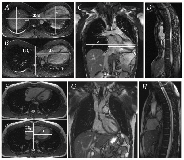
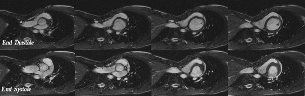
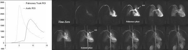
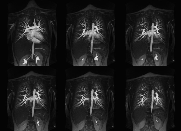
Thirty patients with pectus excavatum deformity (23 men, 7 women, age range: 14-67 years) underwent CMR using 1.5-Tesla scanner (Siemens) and were compared to 25 healthy controls (18 men, 7 women, age range 18-50 years). The CMR protocol included cardiac cine images, pulmonary artery flow quantification, time resolved 3D contrast enhanced MR angiography (CEMRA) and high spatial resolution CEMRA. Chest wall indices including maximum transverse diameter, pectus index (PI), and chest-flatness were measured in all subjects. Left and right ventricular ejection fractions (LVEF, RVEF), ventricular long and short dimensions (LD, SD), mid-ventricle myocardial shortening, pulmonary-systemic circulation time, and pulmonary artery flow were quantified.
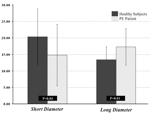
In patients with pectus excavatum, the pectus index was 9.3 ± 5.0 versus 2.8 ± 0.4 in controls (P < 0.001). No significant differences between pectus excavatum patients and controls were found in LV ejection fraction, LV myocardial shortening, pulmonary-systemic circulation time or pulmonary flow indices. In pectus excavatum, resting RV ejection fraction was reduced (53.9 ± 9.6 versus 60.5 ± 9.5; P = 0.013), RVSD was reduced (P < 0.05) both at end diastole and systole, RVLD was increased at end diastole (P < 0.05) reflecting geometric distortion of the RV due to sternal compression.
Depression of the sternum in pectus excavatum patients distorts RV geometry. Resting RVEF was reduced by 6% of the control value, suggesting that these geometrical changes may influence myocardial performance. Resting LV function, pulmonary circulation times and pulmonary vascular anatomy and perfusion indices were no different to controls.
使用心血管磁共振成像(CMR)评估漏斗胸患者与对照者的心胸结构和功能。
30 例漏斗胸畸形患者(男 23 例,女 7 例,年龄 14-67 岁)在 1.5T 磁共振扫描仪(西门子)上进行 CMR 检查,并与 25 名健康对照者(男 18 例,女 7 例,年龄 18-50 岁)进行比较。CMR 方案包括心脏电影图像、肺动脉流量定量、时间分辨 3D 对比增强磁共振血管造影(CEMRA)和高空间分辨率 CEMRA。所有受试者均测量胸廓指数,包括最大横径、胸壁指数(PI)和胸廓平坦度。定量评估左、右心室射血分数(LVEF、RVEF)、心室长、短径(LD、SD)、心室中部心肌缩短率、肺-体循环时间和肺动脉流量。
在漏斗胸患者中,胸壁指数为 9.3 ± 5.0,而对照组为 2.8 ± 0.4(P < 0.001)。漏斗胸患者与对照组之间在左心室射血分数、左心室心肌缩短率、肺-体循环时间或肺血流指数方面无显著差异。在漏斗胸患者中,静息右心室射血分数降低(53.9 ± 9.6 比 60.5 ± 9.5;P = 0.013),右心室短轴舒张末期和收缩末期均减小(P < 0.05),右心室长轴舒张末期增大(P < 0.05),这反映了胸骨压迫导致的右心室几何变形。
漏斗胸患者胸骨凹陷导致右心室几何形状变形。静息右心室射血分数比对照组降低 6%,提示这些几何变化可能影响心肌功能。静息左心室功能、肺循环时间以及肺血管解剖和灌注指数与对照组无差异。