• 文献检索
  • 文档翻译
  • 深度研究
  • 学术资讯
  • Suppr Zotero 插件Zotero 插件
  • 邀请有礼
  • 套餐&价格
  • 历史记录
应用&插件
Suppr Zotero 插件Zotero 插件浏览器插件Mac 客户端Windows 客户端微信小程序
定价
高级版会员购买积分包购买API积分包
服务
文献检索文档翻译深度研究API 文档MCP 服务
关于我们
关于 Suppr公司介绍联系我们用户协议隐私条款
关注我们

Suppr 超能文献

核心技术专利:CN118964589B侵权必究
粤ICP备2023148730 号-1Suppr @ 2026

文献检索

告别复杂PubMed语法,用中文像聊天一样搜索,搜遍4000万医学文献。AI智能推荐,让科研检索更轻松。

立即免费搜索

文件翻译

保留排版,准确专业,支持PDF/Word/PPT等文件格式,支持 12+语言互译。

免费翻译文档

深度研究

AI帮你快速写综述,25分钟生成高质量综述,智能提取关键信息,辅助科研写作。

立即免费体验

一种新的工具,通过半自动图像处理管道评估漏斗胸,计算经典的严重程度指数和一个新的标记:容积矫正指数。

A new tool for assessing Pectus Excavatum by a semi-automatic image processing pipeline calculating the classical severity indexes and a new marker: the Volumetric Correction Index.

机构信息

Department of Informatics, Bioengineering Robotics and System Engineering (DIBRIS), University of Genoa, Viale Causa 13, 16143, Genova, Italy.

Complex Operative Radiology Unit, IRCCS Giannina Gaslini, Genova, Italy.

出版信息

BMC Med Imaging. 2022 Feb 20;22(1):30. doi: 10.1186/s12880-022-00754-0.

DOI:10.1186/s12880-022-00754-0
PMID:35184746
原文链接:https://pmc.ncbi.nlm.nih.gov/articles/PMC8859892/
Abstract

BACKGROUND

In clinical assessment of Pectus Excavatum (PE), the indication to surgery is based not only on symptoms but also on quantitative markers calculated from Computed Tomography (CT) or Magnetic Resonance Imaging (MRI) scans. According to clinical routine, these indexes are measured manually by radiologists with limited computer support. This process is time consuming and potentially subjected to inaccuracy and individual variability in measurements. Moreover, the existing indexes have limitations, since they are based on linear measurements performed on single slices rather than on volumetric data derived from all the thoracic scans.

RESULTS

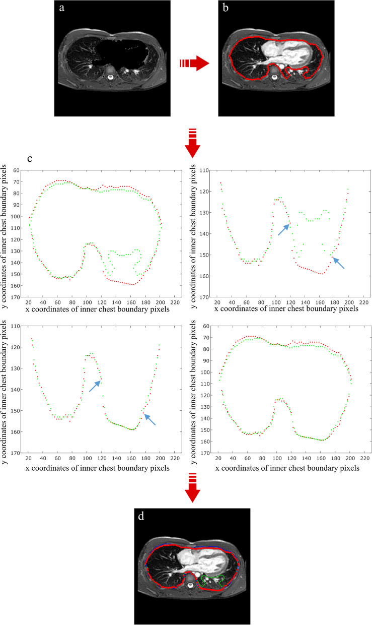
In this paper we present an image processing pipeline aimed at providing radiologists with a computer-aid tool in support of diagnosis of PE patients developed in MATLAB® and conceived for MRI images. This framework has a dual purpose: (i) to automatize computation of clinical indexes with a view to ease and standardize pre-operative evaluation; (ii) to propose a new marker of pathological severity based on volumetric analysis and overcoming the limitations of existing axial slice-based indexes. Final designed framework is semi-automatic, requiring some user interventions at crucial steps: this is realized through a Graphical User Interface (GUI) that simplifies the interaction between the user and the tools. We tested our pipeline on 50 pediatric patients from Gaslini Children's Hospital and performed manual computation of indexes, comparing the results between the proposed tool and gold-standard clinical practice. Automatic indexes provided by our algorithm have shown good agreement with manual measurements by two independent readers. Moreover, the new proposed Volumetric Correction Index (VCI) has exhibited good correlation with standardized markers of pathological severity, proving to be a potential innovative tool for diagnosis, treatment, and follow-up.

CONCLUSIONS

Our pipeline represents an innovative image processing in PE evaluation, based on MRI images (radiation-free) and providing the clinician with a quick and accurate tool for automatically calculating the classical PE severity indexes and a new more comprehensive marker: the Volumetric Correction Index.

摘要

背景

在漏斗胸(PE)的临床评估中,手术指征不仅基于症状,还基于从计算机断层扫描(CT)或磁共振成像(MRI)扫描计算得出的定量指标。根据临床常规,这些指标由放射科医生在有限的计算机支持下手动测量。这个过程既耗时又容易出现不准确和测量的个体差异。此外,现有的指标存在局限性,因为它们基于对单个切片的线性测量,而不是基于从所有胸部扫描得出的容积数据。

结果

在本文中,我们提出了一个图像处理管道,旨在为放射科医生提供一种计算机辅助工具,以支持在 MATLAB®中开发的用于 MRI 图像的 PE 患者的诊断。该框架具有双重目的:(i)自动计算临床指标,以简化和标准化术前评估;(ii)提出一种基于容积分析的新的病理严重程度指标,克服了基于现有轴向切片指标的局限性。最终设计的框架是半自动的,在关键步骤需要一些用户干预:这是通过一个图形用户界面(GUI)实现的,它简化了用户与工具之间的交互。我们在 Gaslini 儿童医院的 50 名儿科患者上测试了我们的管道,并对索引进行了手动计算,比较了我们的工具和黄金标准临床实践的结果。我们算法提供的自动索引与两位独立读者的手动测量结果具有良好的一致性。此外,新提出的容积校正指数(VCI)与病理严重程度的标准化指标具有良好的相关性,证明它是一种用于诊断、治疗和随访的潜在创新工具。

结论

我们的管道代表了基于 MRI 图像(无辐射)的漏斗胸评估中的一种创新的图像处理方法,为临床医生提供了一种快速准确的工具,用于自动计算经典的漏斗胸严重程度指数和一个新的更全面的指标:容积校正指数。

https://cdn.ncbi.nlm.nih.gov/pmc/blobs/8de1/8859892/7535f8cb6a4b/12880_2022_754_Fig7_HTML.jpg
https://cdn.ncbi.nlm.nih.gov/pmc/blobs/8de1/8859892/f88a529e1107/12880_2022_754_Fig1_HTML.jpg
https://cdn.ncbi.nlm.nih.gov/pmc/blobs/8de1/8859892/c3db5261581d/12880_2022_754_Fig2_HTML.jpg
https://cdn.ncbi.nlm.nih.gov/pmc/blobs/8de1/8859892/559dcd511ec4/12880_2022_754_Fig3_HTML.jpg
https://cdn.ncbi.nlm.nih.gov/pmc/blobs/8de1/8859892/659425f908e6/12880_2022_754_Fig4_HTML.jpg
https://cdn.ncbi.nlm.nih.gov/pmc/blobs/8de1/8859892/cbfa19904cb4/12880_2022_754_Fig5_HTML.jpg
https://cdn.ncbi.nlm.nih.gov/pmc/blobs/8de1/8859892/1c41cc5005b2/12880_2022_754_Fig6_HTML.jpg
https://cdn.ncbi.nlm.nih.gov/pmc/blobs/8de1/8859892/7535f8cb6a4b/12880_2022_754_Fig7_HTML.jpg

相似文献

1
A new tool for assessing Pectus Excavatum by a semi-automatic image processing pipeline calculating the classical severity indexes and a new marker: the Volumetric Correction Index.一种新的工具,通过半自动图像处理管道评估漏斗胸,计算经典的严重程度指数和一个新的标记:容积矫正指数。
BMC Med Imaging. 2022 Feb 20;22(1):30. doi: 10.1186/s12880-022-00754-0.
2
Enhancing pectus excavatum diagnosis with an automated batch evaluation tool for chest computed tomography images.利用胸部 CT 图像自动批量评估工具增强漏斗胸的诊断。
Sci Rep. 2024 Oct 8;14(1):23468. doi: 10.1038/s41598-024-73880-0.
3
Comparison of Haller index values calculated with chest radiographs versus CT for pectus excavatum evaluation.比较采用胸部 X 光片和 CT 计算的 Haller 指数值在漏斗胸评估中的应用。
Pediatr Radiol. 2010 Nov;40(11):1763-7. doi: 10.1007/s00247-010-1681-z. Epub 2010 May 15.
4
A computational pipeline for quantification of pulmonary infections in small animal models using serial PET-CT imaging.使用连续 PET-CT 成像对小动物模型中的肺部感染进行定量分析的计算流程。
EJNMMI Res. 2013 Jul 23;3(1):55. doi: 10.1186/2191-219X-3-55.
5
Evaluation of pectus excavatum indexes during standard cardiac magnetic resonance: Potential for single preoperative tool.标准心脏磁共振成像期间漏斗胸指数的评估:作为单一术前工具的潜力。
Clin Imaging. 2019 Jan-Feb;53:138-142. doi: 10.1016/j.clinimag.2018.10.013. Epub 2018 Oct 15.
6
Validating 3D indexes in the non-surgical pectus excavatum patient.验证非手术漏斗胸患者的三维指标。
J Pediatr Surg. 2021 Feb;56(2):230-234. doi: 10.1016/j.jpedsurg.2020.06.006. Epub 2020 Jun 11.
7
Relationship between cardiac MR compression classification and CT chest wall indexes in patients with pectus excavatum.漏斗胸患者心脏磁共振压缩分类与胸部CT胸壁指数的关系
J Pediatr Surg. 2018 Nov;53(11):2294-2298. doi: 10.1016/j.jpedsurg.2018.05.015. Epub 2018 May 23.
8
Measuring the impact of surgical intervention on pediatric pectus excavatum using white light scanning.使用白光扫描测量手术干预对小儿漏斗胸的影响。
J Pediatr Surg. 2019 Nov;54(11):2261-2267. doi: 10.1016/j.jpedsurg.2019.01.007. Epub 2019 Jan 23.
9
Utility of radiation-free imaging for initial evaluation of pectus excavatum.无辐射成像在漏斗胸初始评估中的应用
Interact Cardiovasc Thorac Surg. 2019 Oct 1;29(4):503-509. doi: 10.1093/icvts/ivz145.
10
Automatic Assessment of Pectus Excavatum Severity From CT Images Using Deep Learning.基于深度学习的 CT 图像鸡胸严重程度自动评估。
IEEE J Biomed Health Inform. 2022 Jan;26(1):324-333. doi: 10.1109/JBHI.2021.3090966. Epub 2022 Jan 17.

引用本文的文献

1
Enhancing pectus excavatum diagnosis with an automated batch evaluation tool for chest computed tomography images.利用胸部 CT 图像自动批量评估工具增强漏斗胸的诊断。
Sci Rep. 2024 Oct 8;14(1):23468. doi: 10.1038/s41598-024-73880-0.
2
Pectus excavatum and carinatum: a narrative review of epidemiology, etiopathogenesis, clinical features, and classification.漏斗胸和鸡胸:关于流行病学、病因发病机制、临床特征及分类的叙述性综述
J Thorac Dis. 2024 Feb 29;16(2):1687-1701. doi: 10.21037/jtd-23-957. Epub 2024 Jan 22.
3
Advancements in preoperative imaging of pectus excavatum: a comprehensive review.

本文引用的文献

1
An Initial 5-Year Single-Center Experience of 365 Patients Undergoing the Video-Assisted Thoracoscopic Surgery for Nuss Procedure for Pectus Excavatum in Resource-Scare Setting.在资源匮乏环境下365例漏斗胸患者接受胸腔镜辅助Nuss手术的单中心5年初步经验
Front Surg. 2021 Jun 14;8:693562. doi: 10.3389/fsurg.2021.693562. eCollection 2021.
2
Automatic Assessment of Pectus Excavatum Severity From CT Images Using Deep Learning.基于深度学习的 CT 图像鸡胸严重程度自动评估。
IEEE J Biomed Health Inform. 2022 Jan;26(1):324-333. doi: 10.1109/JBHI.2021.3090966. Epub 2022 Jan 17.
3
Cardiovascular MRI assessment of pectus excavatum in pediatric patients and postoperative simulation using vacuum bell.
漏斗胸术前影像学进展:全面综述
J Thorac Dis. 2024 Jan 30;16(1):696-707. doi: 10.21037/jtd-23-662. Epub 2024 Jan 24.
采用真空钟对小儿漏斗胸患者进行心血管 MRI 评估及术后模拟
J Pediatr Surg. 2021 Sep;56(9):1600-1605. doi: 10.1016/j.jpedsurg.2020.11.017. Epub 2020 Nov 20.
4
Computer-aided diagnosis of pectus excavatum using CT images and deep learning methods.基于 CT 图像和深度学习方法的漏斗胸计算机辅助诊断。
Sci Rep. 2020 Nov 20;10(1):20294. doi: 10.1038/s41598-020-77361-y.
5
Comparison of magnetic resonance imaging and computed tomography to measure preoperative parameters of children with pectus excavatum.磁共振成像与计算机断层扫描在测量漏斗胸患儿术前参数方面的比较。
Pediatr Investig. 2019 Jun 25;3(2):102-109. doi: 10.1002/ped4.12132. eCollection 2019 Jun.
6
Impact of pectus excavatum on cardiac morphology and function according to the site of maximum compression: effect of physical exertion and respiratory cycle.漏斗胸对心脏形态和功能的影响与最大压迫部位有关:体力活动和呼吸周期的影响。
Eur Heart J Cardiovasc Imaging. 2020 Jan 1;21(1):77-84. doi: 10.1093/ehjci/jez061.
7
The prevalence of chest wall deformity in Turkish children.土耳其儿童胸壁畸形的患病率。
Turk J Med Sci. 2018 Dec 12;48(6):1200-1206. doi: 10.3906/sag-1807-180.
8
Evaluation of pectus excavatum indexes during standard cardiac magnetic resonance: Potential for single preoperative tool.标准心脏磁共振成像期间漏斗胸指数的评估:作为单一术前工具的潜力。
Clin Imaging. 2019 Jan-Feb;53:138-142. doi: 10.1016/j.clinimag.2018.10.013. Epub 2018 Oct 15.
9
Relationship between cardiac MR compression classification and CT chest wall indexes in patients with pectus excavatum.漏斗胸患者心脏磁共振压缩分类与胸部CT胸壁指数的关系
J Pediatr Surg. 2018 Nov;53(11):2294-2298. doi: 10.1016/j.jpedsurg.2018.05.015. Epub 2018 May 23.
10
Advantages of Cardiac Magnetic Resonance Imaging for Severe Pectus Excavatum Assessment in Children.心脏磁共振成像在儿童重度漏斗胸评估中的优势
Eur J Pediatr Surg. 2018 Feb;28(1):34-38. doi: 10.1055/s-0037-1604427. Epub 2017 Jul 31.