Gesslein Bodil, Håkansson Gisela, Gustafsson Lotta, Ekström Per, Malmsjö Malin
Department of Ophthalmology, Lund University, Sweden.
Mol Vis. 2010 Nov 6;16:2317-27.
Numerous studies have been performed aimed at limiting the extent of retinal injury after ischemia, but there is still no effective pharmacological treatment available. The aim of the present study was to examine the role of tumor necrosis factor (TNF)α and its receptors (TNF-R1 and TNF-R2), especially considering the neuroretina and the retinal vasculature since the retinal blood vessels are key organs in circulatory failure.
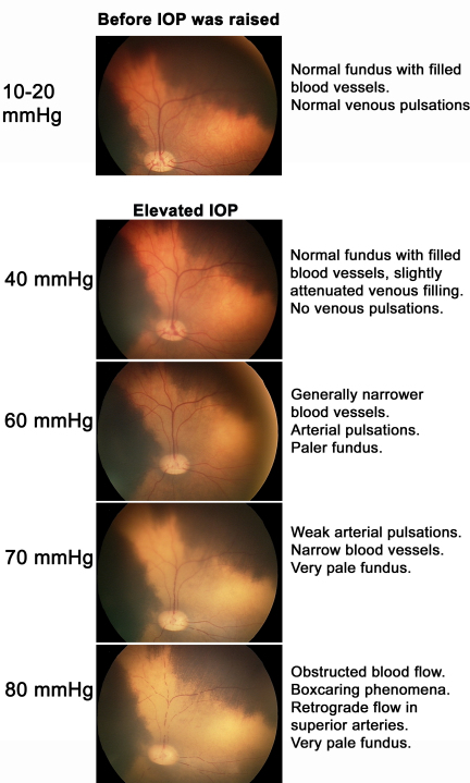
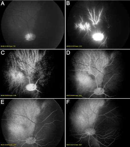
Retinal ischemia was induced in pigs by elevating the intraocular pressure to 80 mmHg in one eye, while the other eye served as a control (sham-operated). One hour of ischemia was followed by 5 or 12 h of reperfusion. Retinal circulation was examined in vivo by fundus imaging and fluorescein angiography. TNF-α levels were measured in the vitreous using an angiogenesis antibody array test. The presence and amounts of TNF-α, TNF-R1, and TNF-R2 were investigated in the neuroretina and in the retinal blood vessels, using immunofluorescence staining and real-time PCR techniques.
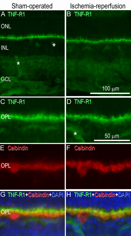
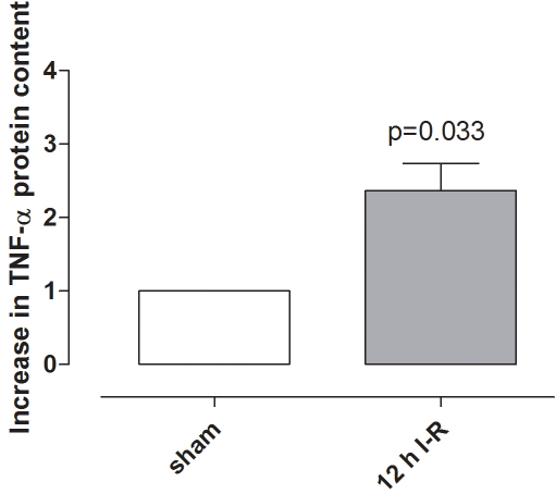
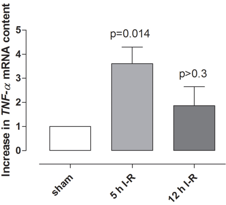
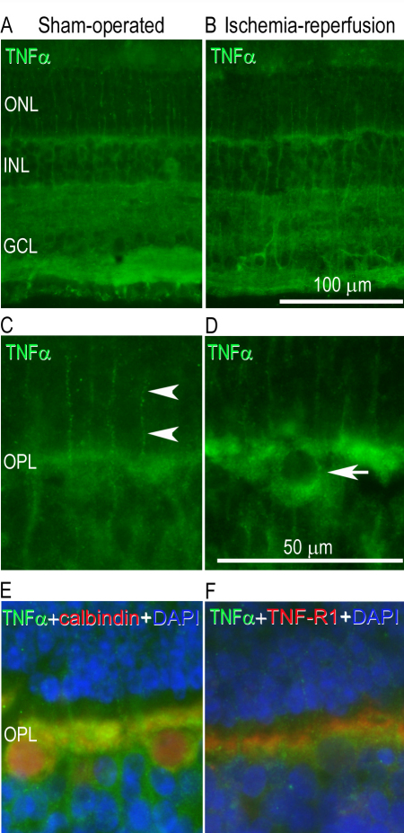
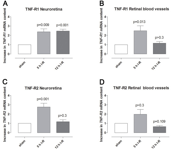
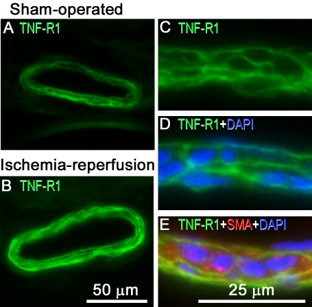
Fundus imaging showed obstructed blood flow when ischemia was induced, and reperfusion was clearly visualized using fluorescein angiography. Ischemia resulted in elevated levels of TNF-α protein in the vitreous and TNF-α mRNA in the neuroretina. TNF-α immunofluorescence staining was localized to the Müller cells and the outer plexiform layer of the neuroretina. The expression of TNF-R1 and TNF-R2 mRNA was increased in both the neuroretina and retinal arteries following ischemia-reperfusion. Immunofluorescence double staining for TNF-R1 and either smooth muscle actin or 4',6-diamidino-2-phenylindole (DAPI) indicated expression in the cell membranes of the vascular smooth muscle cells. Double staining with TNF-R1 and calbindin showed localization to the horizontal cells in the outer plexiform layer of the neuroretina.
Retinal ischemia results in increased expression of TNF-α and its receptors (TNF-R1 and TNF-R2). Cellular signaling pathways involving TNF may be important in the development of retinal injury following ischemia and thus an interesting target for future development of pharmacological therapeutics.
已经进行了大量研究旨在限制缺血后视网膜损伤的程度,但目前仍没有有效的药物治疗方法。本研究的目的是研究肿瘤坏死因子(TNF)α及其受体(TNF-R1和TNF-R2)的作用,特别是考虑到神经视网膜和视网膜血管,因为视网膜血管是循环衰竭中的关键器官。
通过将一只眼睛的眼压升高至80 mmHg诱导猪的视网膜缺血,而另一只眼睛作为对照(假手术)。缺血1小时后进行5或12小时的再灌注。通过眼底成像和荧光素血管造影在体内检查视网膜循环。使用血管生成抗体阵列试验测量玻璃体中的TNF-α水平。使用免疫荧光染色和实时PCR技术研究神经视网膜和视网膜血管中TNF-α、TNF-R1和TNF-R2的存在及含量。
眼底成像显示诱导缺血时血流受阻,荧光素血管造影清晰显示再灌注情况。缺血导致玻璃体中TNF-α蛋白水平升高以及神经视网膜中TNF-α mRNA水平升高。TNF-α免疫荧光染色定位于Müller细胞和神经视网膜的外丛状层。缺血再灌注后,神经视网膜和视网膜动脉中TNF-R1和TNF-R2 mRNA的表达均增加。TNF-R1与平滑肌肌动蛋白或4',6-二脒基-2-苯基吲哚(DAPI)的免疫荧光双重染色表明在血管平滑肌细胞的细胞膜中有表达。TNF-R1与钙结合蛋白的双重染色显示定位于神经视网膜外丛状层的水平细胞。
视网膜缺血导致TNF-α及其受体(TNF-R1和TNF-R2)表达增加。涉及TNF的细胞信号通路在缺血后视网膜损伤的发展中可能很重要,因此是未来药物治疗开发的一个有意义的靶点。