Department of Clinical Sciences, Division of Experimental Vascular Research, Lund University, Lund, Sweden.
J Neuroinflammation. 2011 Aug 28;8:107. doi: 10.1186/1742-2094-8-107.
Tumour necrosis factor-α (TNF-α) is a pleiotropic pro-inflammatory cytokine, which is rapidly upregulated in the brain after injury. TNF-α acts by binding to its receptors, TNF-R1 (p55) and TNF-R2 (p75), on the cell surface. The aim of this study was first to investigate if there is altered expression of TNF-α and TNF-α receptors in cerebral artery walls following global or focal ischemia, and after organ culture. Secondly, we asked if the expression was regulated via activation of the MEK-ERK1/2 pathway.
The hypothesis was tested in vivo after subarachnoid hemorrhage (SAH) and middle cerebral artery occlusion (MCAO), and in vitro by organ culture of isolated cerebral arteries. The localization and amount of TNF-α, TNF-α receptor 1 and 2 proteins were analysed by immunohistochemistry and western blot after 24 and 48 h of organ culture and at 48 h following SAH or MCAO. In addition, cerebral arteries were incubated for 24 or 48 h in the absence or presence of a B-Raf inhibitor (SB386023-b), a MEK- inhibitor (U0126) or an NF-κB inhibitor (IMD-0354), and protein expression evaluated.
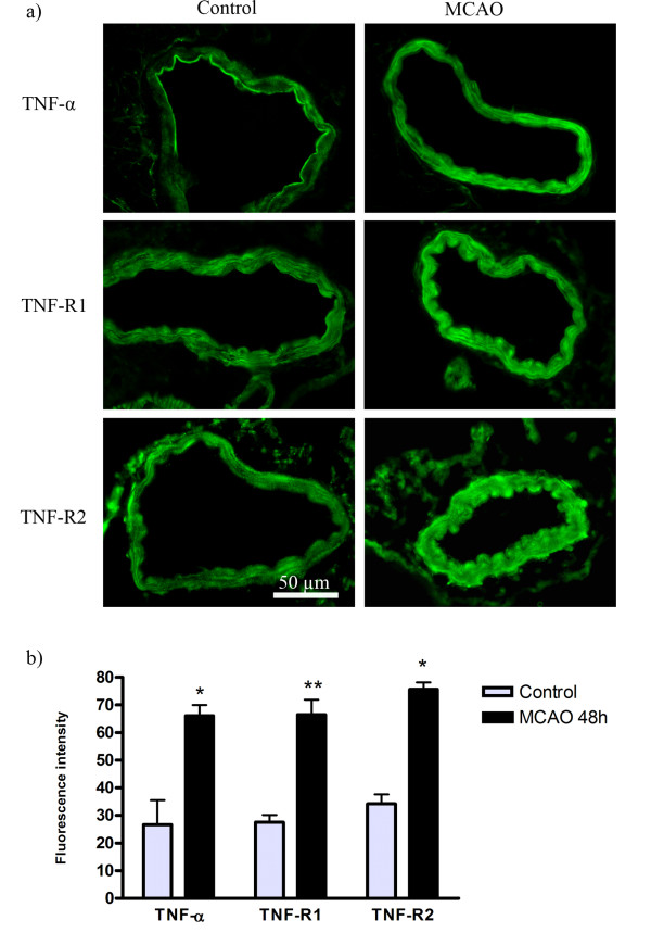
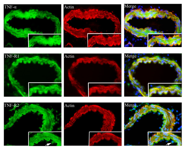
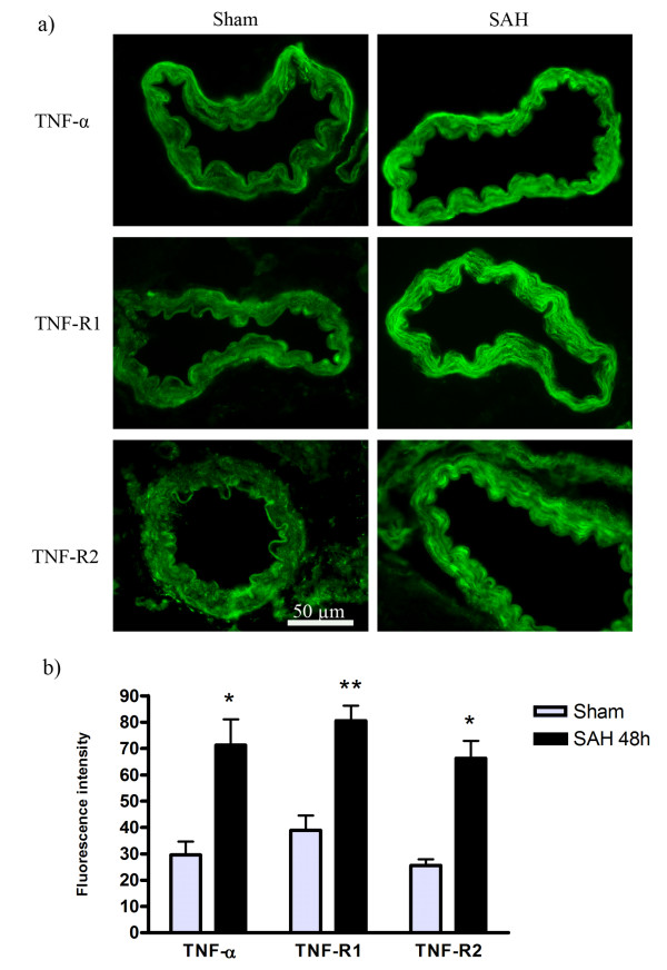
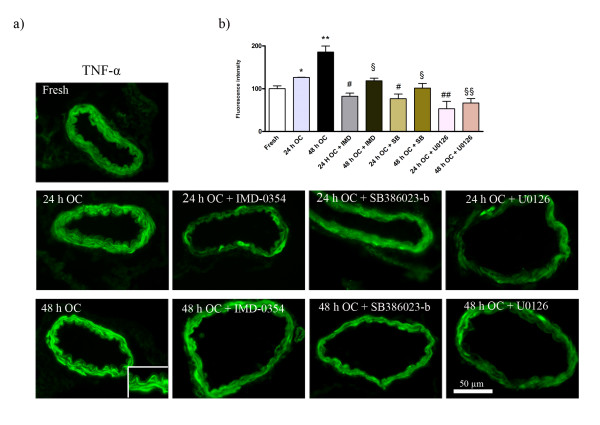
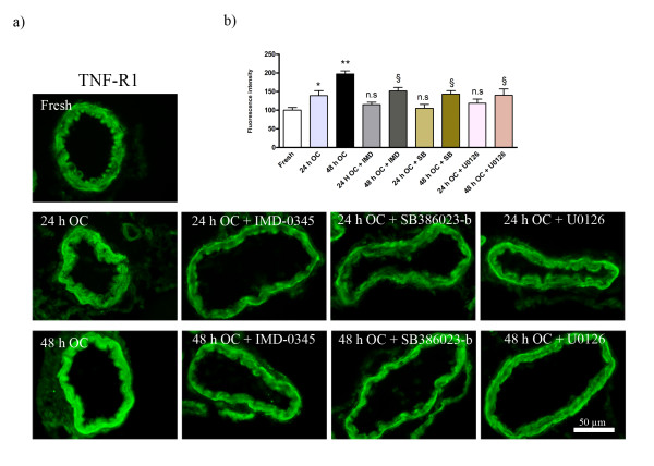
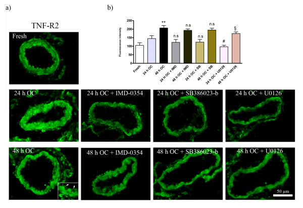
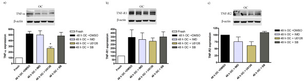
Immunohistochemistry revealed enhanced expression of TNF-α, TNF-R1 and TNF-R2 in the walls of cerebral arteries at 48 h after MCAO and SAH compared with control. Co-localization studies showed that TNF-α, TNF-R1 and TNF-R2 were primarily localized to the cell membrane and the cytoplasm of the smooth muscle cells (SMC). There was, in addition, some expression of TNF-R2 in the endothelial cells. Immunohistochemistry and western blot analysis showed that these proteins were upregulated after 24 and 48 h in culture, and this upregulation reached an apparent maximum at 48 h of organ culture. Treatment with U0126 significantly reduced the enhanced SMC expression of TNF-α, TNF-R1 and TNF-R2 immunoreactivities after 24 and 48 h of organ culture. The Raf and NF-κB inhibitors significantly reduced organ culture induced TNF-α expression while they had minor effects on the TNF-α receptors.
The present study shows that cerebral ischemia and organ culture induce expression of TNF-α and its receptors in the walls of cerebral arteries and that upregulation is transcriptionally regulated via the MEK/ERK pathway.
肿瘤坏死因子-α(TNF-α)是一种多效性促炎细胞因子,在损伤后迅速在大脑中上调。TNF-α通过与其细胞表面受体 TNF-R1(p55)和 TNF-R2(p75)结合而发挥作用。本研究的目的首先是研究在全脑或局灶性缺血以及器官培养后,大脑动脉壁中 TNF-α 和 TNF-α 受体的表达是否发生改变。其次,我们询问这种表达是否通过激活 MEK-ERK1/2 途径来调节。
通过蛛网膜下腔出血(SAH)和大脑中动脉闭塞(MCAO)的体内试验以及离体脑动脉器官培养的体外试验来检验假设。在器官培养 24 小时和 48 小时后以及 SAH 或 MCAO 后 48 小时,通过免疫组织化学和 Western blot 分析 TNF-α、TNF-α 受体 1 和 2 蛋白的定位和数量。此外,将大脑动脉在无或存在 B-Raf 抑制剂(SB386023-b)、MEK 抑制剂(U0126)或 NF-κB 抑制剂(IMD-0354)的情况下孵育 24 小时或 48 小时,并评估蛋白表达。
免疫组织化学显示,与对照组相比,MCAO 和 SAH 后 48 小时大脑动脉壁中 TNF-α、TNF-R1 和 TNF-R2 的表达增强。共定位研究表明,TNF-α、TNF-R1 和 TNF-R2 主要定位于平滑肌细胞(SMC)的细胞膜和细胞质。此外,内皮细胞中也有 TNF-R2 的表达。免疫组织化学和 Western blot 分析显示,这些蛋白在培养 24 小时和 48 小时后上调,并且在器官培养 48 小时时达到明显的最大值。U0126 处理显著降低了器官培养 24 小时和 48 小时后 SMC 表达增强的 TNF-α、TNF-R1 和 TNF-R2 免疫反应性。Raf 和 NF-κB 抑制剂显著降低了器官培养诱导的 TNF-α 表达,而对 TNF-α 受体的影响较小。
本研究表明,脑缺血和器官培养诱导大脑动脉壁中 TNF-α 及其受体的表达,而上调是通过 MEK/ERK 途径转录调控的。