Department of Surgery, University of Colorado Denver, Aurora, Colorado, USA.
Cardiovasc Diabetol. 2010 Dec 16;9:90. doi: 10.1186/1475-2840-9-90.
Endothelial inflammatory responses mediated by Toll-like receptors (TLRs), particularly TLR2 and TLR4, play an important role in atherogenesis. While Type 1 diabetes (T1D) promotes the development and progression of atherosclerosis, the effect of T1D on TLR2/4-mediated inflammatory responses in coronary artery endothelial cells (CAECs) remains unclear.
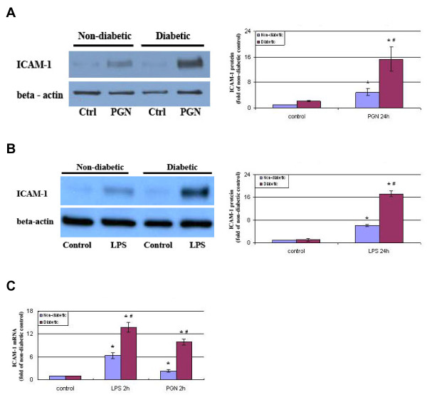
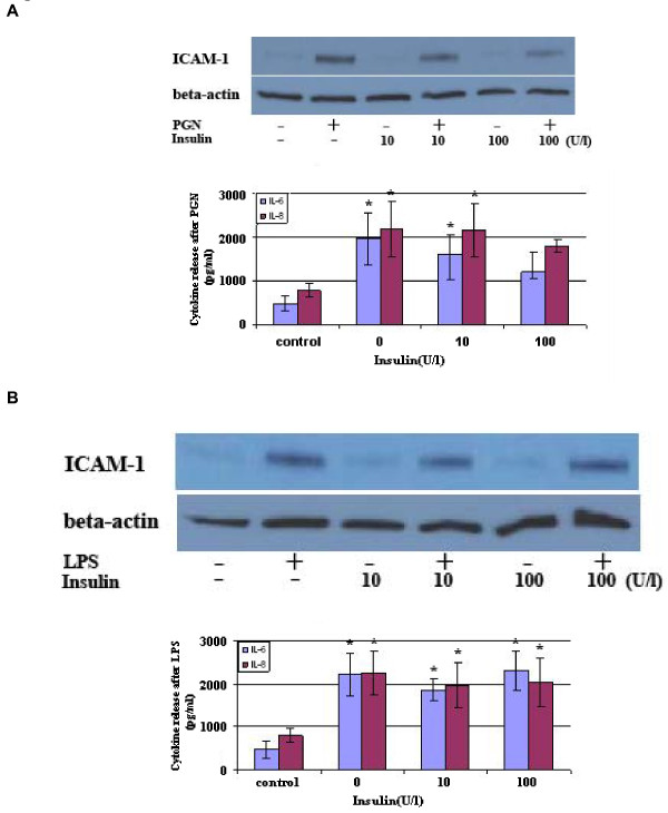
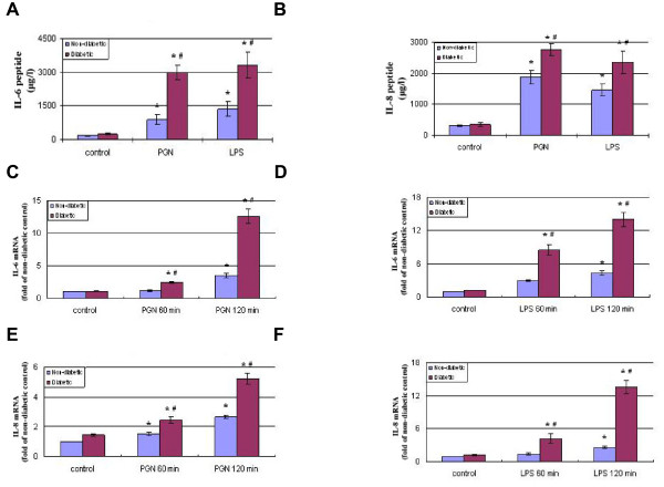
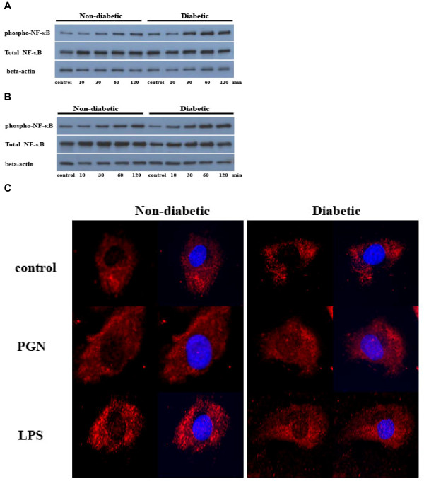
We tested the hypothesis that diabetic CAECs have enhanced inflammatory responses to TLR2/4 stimulation. Non-diabetic and diabetic CAECs were treated with TLR2 agonist peptidoglycan and TLR4 agonist lipopolysaccharide. The expression of ICAM-1, IL-6 and IL-8 were analyzed by real-time PCR, immunoblotting and ELISA, and NF-κB activation by immunoblotting and immunostaining. In additional experiments, insulin was added before TLR stimulation to determine whether insulin deficiency alone is responsible for the alteration of TLR2/4-mediated inflammatory responses.
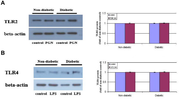
Stimulation of TLR2 or TLR4 induced NF-κB activation, and the expression of ICAM-1, IL-6 and IL-8. Interestingly, the expression of inflammatory mediators was significantly enhanced in diabetic cells. The enhanced inflammatory responses correlated with augmented NF-κB activation in the absence of a change in TLR2 or TLR4 protein levels. Further, pretreatment of diabetic cells with insulin failed to suppress the enhanced inflammatory responses.
Diabetic CAECs have enhanced inflammatory responses to stimulation of TLR2 or TLR4, and insulin alone is insufficient to correct the hyper-inflammatory responses. The mechanism underlying the enhanced inflammatory responses appears to be augmentation of pro-inflammatory signaling, rather than up-regulation of levels of TLR2 and TLR4. These findings suggest that diabetic CAECs adopt a hyper-inflammatory phenotype and that this endothelial phenotypic change may predispose coronary artery to atherogenesis.
Toll 样受体(TLR)介导的内皮炎症反应,尤其是 TLR2 和 TLR4,在动脉粥样硬化的发生发展中起着重要作用。虽然 1 型糖尿病(T1D)促进了动脉粥样硬化的发生和进展,但 T1D 对冠状动脉内皮细胞(CAEC)中 TLR2/4 介导的炎症反应的影响尚不清楚。
我们检验了这样一个假设,即糖尿病 CAEC 对 TLR2/4 刺激的炎症反应增强。用 TLR2 激动剂肽聚糖和 TLR4 激动剂脂多糖处理非糖尿病和糖尿病 CAEC。通过实时 PCR、免疫印迹和 ELISA 分析 ICAM-1、IL-6 和 IL-8 的表达,通过免疫印迹和免疫染色分析 NF-κB 激活。在额外的实验中,在 TLR 刺激前加入胰岛素,以确定胰岛素缺乏是否单独导致 TLR2/4 介导的炎症反应改变。
TLR2 或 TLR4 的刺激诱导 NF-κB 激活和 ICAM-1、IL-6 和 IL-8 的表达。有趣的是,在糖尿病细胞中,炎症介质的表达显著增强。在 TLR2 或 TLR4 蛋白水平没有变化的情况下,增强的炎症反应与 NF-κB 激活增强相关。此外,用胰岛素预处理糖尿病细胞未能抑制增强的炎症反应。
糖尿病 CAEC 对 TLR2 或 TLR4 的刺激有增强的炎症反应,而胰岛素本身不足以纠正过度炎症反应。增强炎症反应的机制似乎是促炎信号的增强,而不是 TLR2 和 TLR4 水平的上调。这些发现表明,糖尿病 CAEC 具有增强的炎症表型,这种内皮表型的改变可能使冠状动脉易发生动脉粥样硬化。