Walberer Maureen, Rueger Maria A, Simard Marie-Lune, Emig Beata, Jander Sebastian, Fink Gereon R, Schroeter Michael
Department of Neurology, University Hospital, Cologne, Germany.
Exp Transl Stroke Med. 2010 Dec 20;2(1):22. doi: 10.1186/2040-7378-2-22.
Neuroinflammation evolves as a multi-facetted response to focal cerebral ischemia. It involves activation of resident glia cell populations, recruitment of blood-derived leucocytes as well as humoral responses. Among these processes, phagocyte accumulation has been suggested to be a surrogate marker of neuroinflammation. We previously assessed phagocyte accumulation in human stroke by MRI. We hypothesize that phagocyte accumulation in the macrosphere model may resemble the temporal and spatial patterns observed in human stroke.
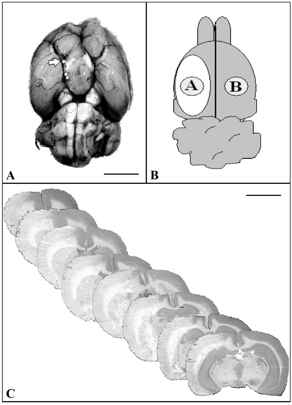
In a rat model of permanent focal ischemia by embolisation of TiO2-spheres we assessed key features of post-ischemic neuroinflammation by the means of histology, immunocytochemistry of glial activation and influx of hematogeneous cells, and quantitative PCR of TNF-α, IL-1, IL-18, and iNOS mRNA.
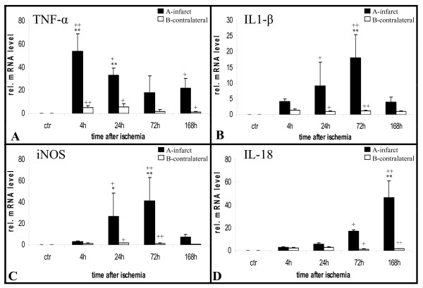
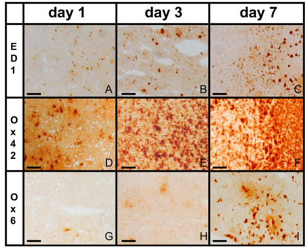
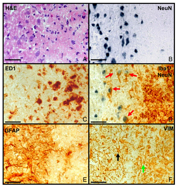
In the boundary zone of the infarct, a transition of ramified microglia into ameboid phagocytic microglia was accompanied by an up-regulation of MHC class II on the cells after 3 days. By day 7, a hypercellular infiltrate consisting of activated microglia and phagocytic cells formed a thick rim around the ischemic infarct core. Interestingly, in the ischemic core microglia could only be observed at day 7. TNF-α was induced rapidly within hours, IL-1β and iNOS peaked within days, and IL-18 later at around 1 week after ischemia.
The macrosphere model closely resembles the characteristical dynamics of postischemic inflammation previously observed in human stroke. We therefore suggest that the macrosphere model is highly appropriate for studying the pathophysiology of stroke in a translational approach from rodent to human.
神经炎症是对局灶性脑缺血的多方面反应。它涉及常驻神经胶质细胞群的激活、血液来源白细胞的募集以及体液反应。在这些过程中,吞噬细胞的积累被认为是神经炎症的替代标志物。我们之前通过磁共振成像评估了人类中风中吞噬细胞的积累情况。我们假设在微球模型中吞噬细胞的积累可能类似于在人类中风中观察到的时空模式。
在通过二氧化钛球栓塞建立的大鼠永久性局灶性缺血模型中,我们通过组织学、胶质细胞激活和造血细胞流入的免疫细胞化学以及肿瘤坏死因子-α、白细胞介素-1、白细胞介素-18和诱导型一氧化氮合酶mRNA的定量聚合酶链反应来评估缺血后神经炎症的关键特征。
在梗死灶的边界区,分支状小胶质细胞向阿米巴样吞噬性小胶质细胞的转变在3天后伴随着细胞上主要组织相容性复合体II类的上调。到第7天,由活化的小胶质细胞和吞噬细胞组成的细胞浸润在缺血梗死核心周围形成了一个厚边缘。有趣的是,在缺血核心中仅在第7天观察到小胶质细胞。肿瘤坏死因子-α在数小时内迅速诱导,白细胞介素-1β和诱导型一氧化氮合酶在数天内达到峰值,白细胞介素-18在缺血后约1周时达到峰值。
微球模型与先前在人类中风中观察到的缺血后炎症的特征性动态非常相似。因此,我们认为微球模型非常适合以从啮齿动物到人类的转化方法研究中风的病理生理学。