• 文献检索
  • 文档翻译
  • 深度研究
  • 学术资讯
  • Suppr Zotero 插件Zotero 插件
  • 邀请有礼
  • 套餐&价格
  • 历史记录
应用&插件
Suppr Zotero 插件Zotero 插件浏览器插件Mac 客户端Windows 客户端微信小程序
定价
高级版会员购买积分包购买API积分包
服务
文献检索文档翻译深度研究API 文档MCP 服务
关于我们
关于 Suppr公司介绍联系我们用户协议隐私条款
关注我们

Suppr 超能文献

核心技术专利:CN118964589B侵权必究
粤ICP备2023148730 号-1Suppr @ 2026

文献检索

告别复杂PubMed语法,用中文像聊天一样搜索,搜遍4000万医学文献。AI智能推荐,让科研检索更轻松。

立即免费搜索

文件翻译

保留排版,准确专业,支持PDF/Word/PPT等文件格式,支持 12+语言互译。

免费翻译文档

深度研究

AI帮你快速写综述,25分钟生成高质量综述,智能提取关键信息,辅助科研写作。

立即免费体验

基于血氧依赖的脑磁共振成像造影。

Brain magnetic resonance imaging with contrast dependent on blood oxygenation.

作者信息

Ogawa S, Lee T M, Kay A R, Tank D W

机构信息

Biophysics Research Department, AT&T Bell Laboratories, Murray Hill, NJ 07974.

出版信息

Proc Natl Acad Sci U S A. 1990 Dec;87(24):9868-72. doi: 10.1073/pnas.87.24.9868.

DOI:10.1073/pnas.87.24.9868
PMID:2124706
原文链接:https://pmc.ncbi.nlm.nih.gov/articles/PMC55275/
Abstract

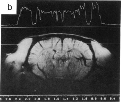
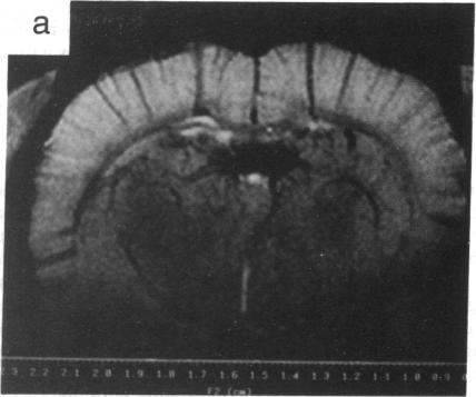
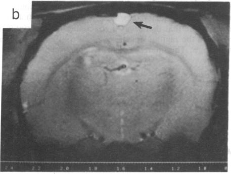
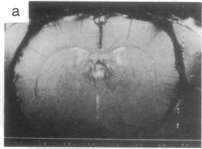
Paramagnetic deoxyhemoglobin in venous blood is a naturally occurring contrast agent for magnetic resonance imaging (MRI). By accentuating the effects of this agent through the use of gradient-echo techniques in high fields, we demonstrate in vivo images of brain microvasculature with image contrast reflecting the blood oxygen level. This blood oxygenation level-dependent (BOLD) contrast follows blood oxygen changes induced by anesthetics, by insulin-induced hypoglycemia, and by inhaled gas mixtures that alter metabolic demand or blood flow. The results suggest that BOLD contrast can be used to provide in vivo real-time maps of blood oxygenation in the brain under normal physiological conditions. BOLD contrast adds an additional feature to magnetic resonance imaging and complements other techniques that are attempting to provide positron emission tomography-like measurements related to regional neural activity.

摘要

静脉血中的顺磁性脱氧血红蛋白是一种天然存在的磁共振成像(MRI)造影剂。通过在高场中使用梯度回波技术增强这种造影剂的效果,我们展示了脑微血管系统的体内图像,其图像对比度反映了血氧水平。这种血氧水平依赖(BOLD)对比度随麻醉剂、胰岛素诱导的低血糖以及改变代谢需求或血流的吸入气体混合物所引起的血氧变化而变化。结果表明,BOLD对比度可用于在正常生理条件下提供脑内血氧合的体内实时图谱。BOLD对比度为磁共振成像增添了一个额外的特性,并补充了其他试图提供与区域神经活动相关的正电子发射断层扫描样测量的技术。

https://cdn.ncbi.nlm.nih.gov/pmc/blobs/3a6f/55275/f0f537f13bac/pnas01049-0373-a.jpg
https://cdn.ncbi.nlm.nih.gov/pmc/blobs/3a6f/55275/2845e2aacfad/pnas01049-0371-a.jpg
https://cdn.ncbi.nlm.nih.gov/pmc/blobs/3a6f/55275/19da470ed0f5/pnas01049-0371-b.jpg
https://cdn.ncbi.nlm.nih.gov/pmc/blobs/3a6f/55275/f3f5cdb3cf73/pnas01049-0371-c.jpg
https://cdn.ncbi.nlm.nih.gov/pmc/blobs/3a6f/55275/b4ec416ecb15/pnas01049-0371-d.jpg
https://cdn.ncbi.nlm.nih.gov/pmc/blobs/3a6f/55275/7b7b847fb8d3/pnas01049-0371-e.jpg
https://cdn.ncbi.nlm.nih.gov/pmc/blobs/3a6f/55275/9a4a3600b4b1/pnas01049-0372-a.jpg
https://cdn.ncbi.nlm.nih.gov/pmc/blobs/3a6f/55275/06ea1e196187/pnas01049-0372-b.jpg
https://cdn.ncbi.nlm.nih.gov/pmc/blobs/3a6f/55275/cd7e09ba91c2/pnas01049-0372-c.jpg
https://cdn.ncbi.nlm.nih.gov/pmc/blobs/3a6f/55275/f0f537f13bac/pnas01049-0373-a.jpg

相似文献

1
Brain magnetic resonance imaging with contrast dependent on blood oxygenation.基于血氧依赖的脑磁共振成像造影。
Proc Natl Acad Sci U S A. 1990 Dec;87(24):9868-72. doi: 10.1073/pnas.87.24.9868.
2
Quantitative magnetic resonance imaging in experimental hypercapnia: improvement in the relation between changes in brain R2 and the oxygen saturation of venous blood after correction for changes in cerebral blood volume.实验性高碳酸血症中的定量磁共振成像:校正脑血容量变化后,脑R2变化与静脉血氧饱和度之间关系的改善。
J Cereb Blood Flow Metab. 1999 Aug;19(8):853-62. doi: 10.1097/00004647-199908000-00004.
3
The sensitivity of magnetic resonance image signals of a rat brain to changes in the cerebral venous blood oxygenation.大鼠大脑磁共振图像信号对脑静脉血氧合变化的敏感性。
Magn Reson Med. 1993 Feb;29(2):205-10. doi: 10.1002/mrm.1910290208.
4
Changes in cerebral blood flow and cerebral oxygen metabolism during neural activation measured by positron emission tomography: comparison with blood oxygenation level-dependent contrast measured by functional magnetic resonance imaging.通过正电子发射断层扫描测量神经激活期间脑血流和脑氧代谢的变化:与功能磁共振成像测量的血氧水平依赖对比的比较。
J Cereb Blood Flow Metab. 2005 Mar;25(3):371-7. doi: 10.1038/sj.jcbfm.9600030.
5
Vascular component analysis of hyperoxic and hypercapnic BOLD contrast.高氧和高碳酸 BOLD 对比的血管成分分析。
Neuroimage. 2012 Feb 1;59(3):2401-12. doi: 10.1016/j.neuroimage.2011.08.110. Epub 2011 Sep 18.
6
Blood-oxygen-level-dependent magnetic resonance signal and cerebral oxygenation responses to brain activation are enhanced by concurrent transient hypertension in rats.大鼠同时发生短暂性高血压时,血氧水平依赖的磁共振信号及脑氧合对脑激活的反应增强。
J Cereb Blood Flow Metab. 2007 Jun;27(6):1280-9. doi: 10.1038/sj.jcbfm.9600436. Epub 2006 Dec 27.
7
Functional brain mapping by blood oxygenation level-dependent contrast magnetic resonance imaging. A comparison of signal characteristics with a biophysical model.基于血氧水平依赖对比的磁共振成像进行脑功能图谱绘制:信号特征与生物物理模型的比较
Biophys J. 1993 Mar;64(3):803-12. doi: 10.1016/S0006-3495(93)81441-3.
8
High-resolution CMR(O2) mapping in rat cortex: a multiparametric approach to calibration of BOLD image contrast at 7 Tesla.大鼠皮层的高分辨率CMR(O2)映射:一种在7特斯拉校准BOLD图像对比度的多参数方法。
J Cereb Blood Flow Metab. 2000 May;20(5):847-60. doi: 10.1097/00004647-200005000-00012.
9
Characterization of cerebrovascular responses to hyperoxia and hypercapnia using MRI in rat.使用MRI对大鼠脑血管对高氧和高碳酸血症的反应进行表征。
Neuroimage. 2009 May 1;45(4):1126-34. doi: 10.1016/j.neuroimage.2008.11.037. Epub 2008 Dec 14.
10
Dynamic magnetic resonance imaging of human brain activity during primary sensory stimulation.初级感觉刺激期间人脑活动的动态磁共振成像
Proc Natl Acad Sci U S A. 1992 Jun 15;89(12):5675-9. doi: 10.1073/pnas.89.12.5675.

引用本文的文献

1
A unified approach for identifying PET-based neuronal activation and molecular connectivity with the functional PET toolbox.一种使用功能性正电子发射断层扫描(PET)工具箱识别基于PET的神经元激活和分子连接性的统一方法。
J Cereb Blood Flow Metab. 2025 Sep 8:271678X251370831. doi: 10.1177/0271678X251370831.
2
SNAKE: A modular realistic fMRI data simulator from the space-time domain to k-space and back.SNAKE:一种从时空域到k空间再返回的模块化逼真功能磁共振成像数据模拟器。
Imaging Neurosci (Camb). 2025 Sep 2;3. doi: 10.1162/IMAG.a.121. eCollection 2025.
3
Decrease and recovery of integrated information Φ during anesthesia and sleep on human functional magnetic resonance imaging.

本文引用的文献

1
NMR chemical shift imaging in three dimensions.三维核磁共振化学位移成像
Proc Natl Acad Sci U S A. 1982 Jun;79(11):3523-6. doi: 10.1073/pnas.79.11.3523.
2
Oxygenation dependence of the transverse relaxation time of water protons in whole blood at high field.高场强下全血中水质子横向弛豫时间的氧合依赖性
Biochim Biophys Acta. 1982 Feb 2;714(2):265-70. doi: 10.1016/0304-4165(82)90333-6.
3
Spatially localized 1H NMR spectra of metabolites in the human brain.人脑代谢物的空间定位1H核磁共振谱。
人类功能磁共振成像中麻醉和睡眠期间整合信息Φ的减少与恢复
Neurosci Conscious. 2025 Sep 1;2025(1):niaf024. doi: 10.1093/nc/niaf024. eCollection 2025.
4
Cortical modulation of resting state BOLD signals in white matter.白质中静息态脑血氧水平依赖信号的皮质调制
Sci Rep. 2025 Aug 17;15(1):30056. doi: 10.1038/s41598-025-14352-x.
5
Quantification of brain functional connectivity deviations in individuals: A scoping review of functional MRI studies.个体脑功能连接偏差的量化:功能磁共振成像研究的范围综述
Neuroimage Clin. 2025 Aug 13;48:103856. doi: 10.1016/j.nicl.2025.103856.
6
Assessing and Improving the Reproducibility of Cerebrovascular Reactivity Evaluations in Healthy Subjects Using Short-Breath-Hold fMRI.使用短屏气功能磁共振成像评估和提高健康受试者脑血管反应性评估的可重复性
Diagnostics (Basel). 2025 Aug 3;15(15):1946. doi: 10.3390/diagnostics15151946.
7
Blood nulling versus tissue suppression: Enhancing integrated VASO and perfusion (VAPER) contrast for laminar fMRI.血液消除与组织抑制:增强用于层流功能磁共振成像的综合血管造影和灌注(VAPER)对比。
Imaging Neurosci (Camb). 2025 Jan 21;3. doi: 10.1162/imag_a_00453. eCollection 2025.
8
Factorized binary search: Change point detection in the network structure of multivariate high-dimensional time series.因式分解二分搜索:多元高维时间序列网络结构中的变化点检测
Imaging Neurosci (Camb). 2025 Apr 17;3. doi: 10.1162/imag_a_00520. eCollection 2025.
9
Somatosensory-evoked response induces extensive diffusivity and kurtosis changes associated with neural activity in rodents.体感诱发电位在啮齿动物中诱发与神经活动相关的广泛扩散率和峰度变化。
Imaging Neurosci (Camb). 2025 Feb 18;3. doi: 10.1162/imag_a_00445. eCollection 2025.
10
Macrovascular contributions to resting-state fMRI signals: A comparison between EPI and bSSFP at 9.4 Tesla.大血管对静息态功能磁共振成像信号的贡献:9.4特斯拉下回波平面成像(EPI)与稳态自由进动序列(bSSFP)的比较
Imaging Neurosci (Camb). 2025 Jan 7;3. doi: 10.1162/imag_a_00435. eCollection 2025.
Proc Natl Acad Sci U S A. 1988 Mar;85(6):1821-5. doi: 10.1073/pnas.85.6.1821.
4
Magnetic resonance imaging of perfusion.灌注的磁共振成像
Magn Reson Med. 1990 May;14(2):283-92. doi: 10.1002/mrm.1910140213.
5
Quantification of regional blood flow by monitoring of exogenous tracer via nuclear magnetic resonance spectroscopy.通过核磁共振波谱监测外源性示踪剂对局部血流进行定量分析。
Magn Reson Med. 1990 May;14(2):266-82. doi: 10.1002/mrm.1910140212.
6
Perfusion imaging with NMR contrast agents.使用核磁共振造影剂的灌注成像。
Magn Reson Med. 1990 May;14(2):249-65. doi: 10.1002/mrm.1910140211.
7
Optimizing blood vessel contrast in fast three-dimensional MRI.在快速三维磁共振成像中优化血管对比度
Magn Reson Med. 1990 May;14(2):202-21. doi: 10.1002/mrm.1910140207.
8
Time-of-flight effects in MR imaging of flow.磁共振血流成像中的飞行时间效应。
Magn Reson Med. 1990 May;14(2):187-93. doi: 10.1002/mrm.1910140205.
9
SMRM Workshop on Magnetic Resonance Imaging of Blood Flow. March 13-14, 1989, Philadelphia, Pennsylvania.磁共振血流成像研讨会。1989年3月13日至14日,宾夕法尼亚州费城
Magn Reson Med. 1990 May;14(2):171-320.
10
Magnetic resonance imaging of blood vessels at high fields: in vivo and in vitro measurements and image simulation.高场强下血管的磁共振成像:体内和体外测量及图像模拟
Magn Reson Med. 1990 Oct;16(1):9-18. doi: 10.1002/mrm.1910160103.