Department of Biomedical Engineering, Case Western Reserve University, Cleveland, Ohio 44106 OH, USA.
Urology. 2011 May;77(5):1254-8. doi: 10.1016/j.urology.2010.11.044. Epub 2011 Jan 22.
To verify the ability to identify the layered structures of the ureteral wall and to image a segment of the ureter in 3 dimensions with high-speed, endoscopic optical coherence tomography (EOCT).
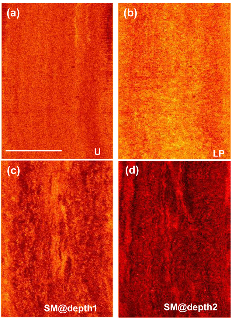
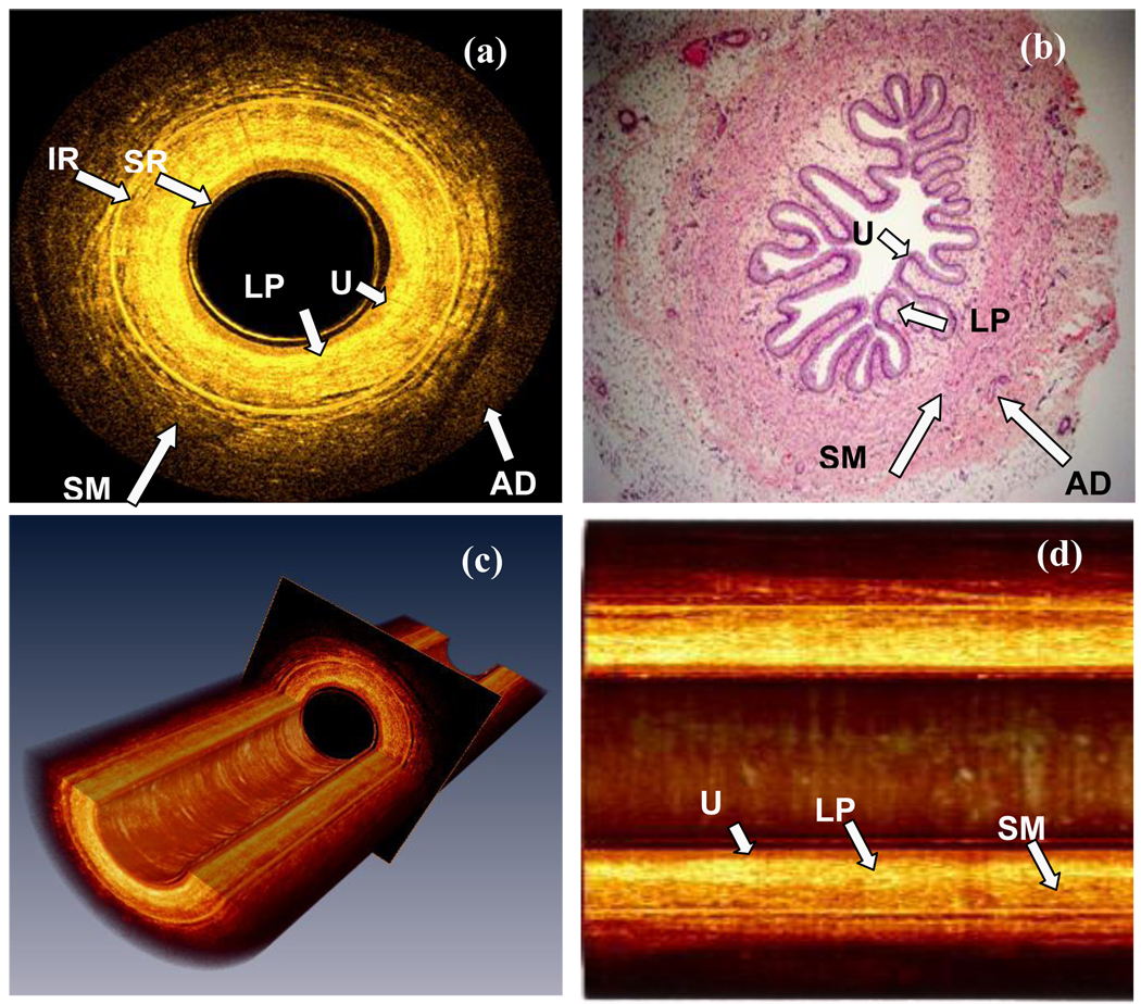
We imaged a porcine ureter ex vivo using a spectral-domain EOCT with a specially designed circumferential scanning fiber catheter. The images were correlated with the histologic findings to identify the corresponding structures. Three-dimensional images and en face images at different depths from the luminal surface were reconstructed from the multiple cross-sectional images to visualize the layered structure of a segment of the ureter from different perspectives.
The EOCT images clearly revealed all layers of the ureteral wall as shown in the histologic images. In particular, with the specially designed fiber catheter, the light beam was well centered during the rotation and pull back, allowing constant acquisition of high-fidelity images and unambiguous identification of the smooth muscle layers in all images. With high-speed EOCT, a segment of ureter (20 mm) can be imaged in <90 seconds at a high resolution.
With its ability to visualize all layers of the ureteral wall, EOCT offers the potential to stage urothelial cancers that have infiltrated the muscular wall (Stage T2). This information will be complimentary to the diagnostic information obtained through ureteroscopic biopsy and computed tomography urogram.
验证高速内窥镜光学相干断层扫描(EOCT)识别输尿管壁分层结构和三维成像输尿管节段的能力。
我们使用专门设计的环形扫描纤维导管对离体猪输尿管进行光谱域 EOCT 成像。将图像与组织学发现相关联,以识别相应的结构。从多个横截面图像重建三维图像和不同深度的表面图像,从不同角度可视化输尿管节段的分层结构。
EOCT 图像清晰地显示了输尿管壁的所有层,与组织学图像一致。特别是,使用专门设计的纤维导管,在旋转和回拉过程中光束能够很好地居中,从而能够持续获取高保真图像,并在所有图像中明确识别平滑肌层。使用高速 EOCT,可在<90 秒内以高分辨率对 20mm 长的输尿管段进行成像。
EOCT 能够可视化输尿管壁的所有层,有望对已浸润肌层的尿路上皮癌(T2 期)进行分期。这些信息将与通过输尿管镜活检和计算机断层尿路造影获得的诊断信息互补。