Neuroimaging Research Unit, Scientific Institute and University Ospedale San Raffaele, Milan, Italy.
PLoS One. 2011 Feb 10;6(2):e17081. doi: 10.1371/journal.pone.0017081.
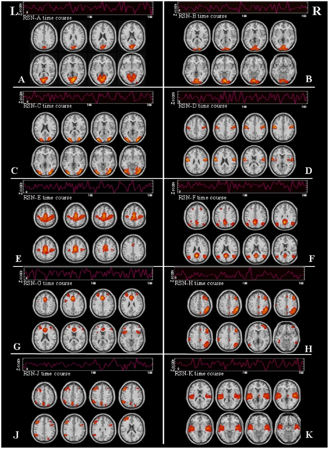
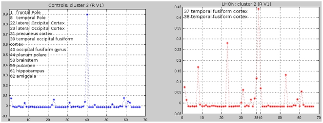
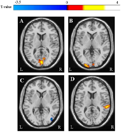
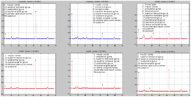
We assessed abnormalities within the principal brain resting state networks (RSNs) in patients with Leber's hereditary optic neuropathy (LHON) to define whether functional abnormalities in this disease are limited to the visual system or, conversely, tend to be more diffuse. We also defined the structural substrates of fMRI changes using a connectivity-based analysis of diffusion tensor (DT) MRI data. Neuro-ophthalmologic assessment, DT MRI and RS fMRI data were acquired from 13 LHON patients and 13 healthy controls. RS fMRI data were analyzed using independent component analysis and SPM5. A DT MRI connectivity-based parcellation analysis was performed using the primary visual and auditory cortices, bilaterally, as seed regions. Compared to controls, LHON patients had a significant increase of RS fluctuations in the primary visual and auditory cortices, bilaterally. They also showed decreased RS fluctuations in the right lateral occipital cortex and right temporal occipital fusiform cortex. Abnormalities of RS fluctuations were correlated significantly with retinal damage and disease duration. The DT MRI connectivity-based parcellation identified a higher number of clusters in the right auditory cortex in LHON vs. controls. Differences of cluster-centroid profiles were found between the two groups for all the four seeds analyzed. For three of these areas, a correspondence was found between abnormalities of functional and structural connectivities. These results suggest that functional and structural abnormalities extend beyond the visual network in LHON patients. Such abnormalities also involve the auditory network, thus corroborating the notion of a cross-modal plasticity between these sensory modalities in patients with severe visual deficits.
我们评估了 Leber 遗传性视神经病变 (LHON) 患者主要脑静息态网络 (RSN) 内的异常,以确定该疾病的功能异常是否仅限于视觉系统,或者相反,是否倾向于更为弥散。我们还使用基于弥散张量 (DT) MRI 数据的连接分析来定义 fMRI 变化的结构基础。从 13 名 LHON 患者和 13 名健康对照中获取了神经眼科评估、DT MRI 和 RS fMRI 数据。使用独立成分分析和 SPM5 分析 RS fMRI 数据。使用双侧初级视觉和听觉皮层作为种子区域,进行基于 DT MRI 连接的分区分析。与对照组相比,LHON 患者双侧初级视觉和听觉皮层的 RS 波动显著增加。他们还显示右侧外侧枕叶和右侧颞枕梭状回的 RS 波动减少。RS 波动的异常与视网膜损伤和疾病持续时间显著相关。基于 DT MRI 连接的分区在 LHON 中比对照组在右侧听觉皮层中识别出更多的聚类。两组之间的所有四个种子分析的聚类质心图谱都存在差异。对于这三个区域,在功能和结构连接的异常之间发现了对应关系。这些结果表明,在 LHON 患者中,功能和结构异常不仅局限于视觉网络。这些异常还涉及听觉网络,从而证实了在严重视力缺陷患者中这些感觉模态之间存在跨模态可塑性的观点。