Harrison Ben J, Pujol Jesus, Ortiz Hector, Fornito Alex, Pantelis Christos, Yücel Murat
Melbourne Neuropsychiatry Centre, Department of Psychiatry, The University of Melbourne and Melbourne Health, Victoria, Australia.
PLoS One. 2008 Mar 19;3(3):e1794. doi: 10.1371/journal.pone.0001794.
There is growing interest in the nature of slow variations of the blood oxygen level-dependent (BOLD) signal observed in functional MRI resting-state studies. In humans, these slow BOLD variations are thought to reflect an underlying or intrinsic form of brain functional connectivity in discrete neuroanatomical systems. While these 'resting-state networks' may be relatively enduring phenomena, other evidence suggest that dynamic changes in their functional connectivity may also emerge depending on the brain state of subjects during scanning.
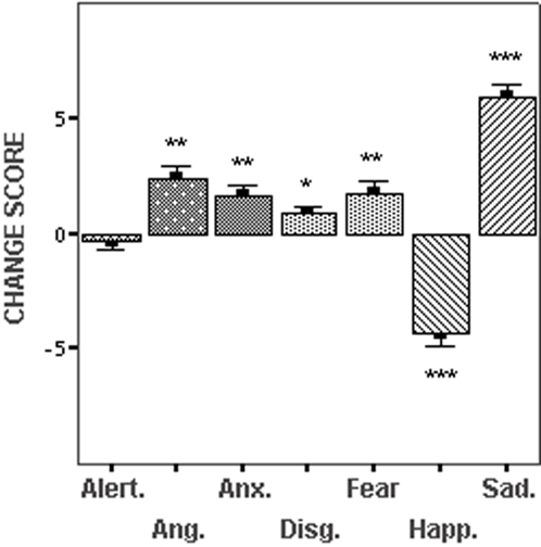
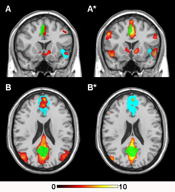
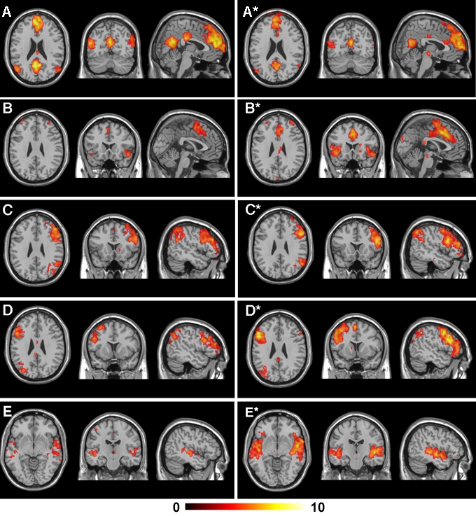
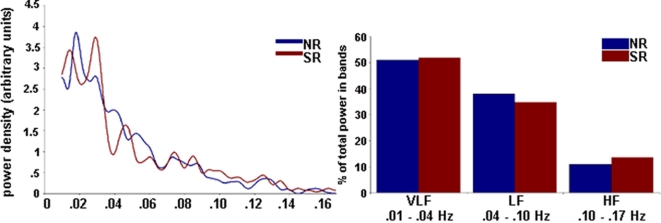
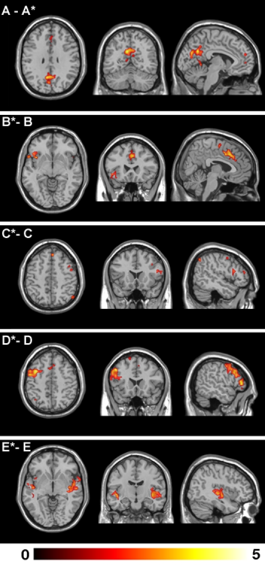
METHODOLOGY/PRINCIPAL FINDINGS: In this study, we examined healthy subjects (n = 24) with a mood induction paradigm during two continuous fMRI recordings to assess the effects of a change in self-generated mood state (neutral to sad) on the functional connectivity of these resting-state networks (n = 24). Using independent component analysis, we identified five networks that were common to both experimental states, each showing dominant signal fluctuations in the very low frequency domain (approximately 0.04 Hz). Between the two states, we observed apparent increases and decreases in the overall functional connectivity of these networks. Primary findings included increased connectivity strength of a paralimbic network involving the dorsal anterior cingulate and anterior insula cortices with subjects' increasing sadness and decreased functional connectivity of the 'default mode network'.
CONCLUSIONS/SIGNIFICANCE: These findings support recent studies that suggest the functional connectivity of certain resting-state networks may, in part, reflect a dynamic image of the current brain state. In our study, this was linked to changes in subjective mood.
在功能磁共振成像静息态研究中,人们对血氧水平依赖(BOLD)信号的缓慢变化性质越来越感兴趣。在人类中,这些缓慢的BOLD变化被认为反映了离散神经解剖系统中潜在的或内在的脑功能连接形式。虽然这些“静息态网络”可能是相对持久的现象,但其他证据表明,根据扫描过程中受试者的脑状态,其功能连接的动态变化也可能出现。
方法/主要发现:在本研究中,我们在两次连续的功能磁共振成像记录期间,使用情绪诱导范式对健康受试者(n = 24)进行了检查,以评估自我产生的情绪状态变化(从中性到悲伤)对这些静息态网络(n = 24)功能连接的影响。使用独立成分分析,我们识别出两种实验状态共有的五个网络,每个网络在极低频域(约0.04 Hz)均显示出占主导地位的信号波动。在两种状态之间,我们观察到这些网络的整体功能连接明显增加和减少。主要发现包括,随着受试者悲伤情绪增加,涉及背侧前扣带回和前岛叶皮质的边缘旁网络的连接强度增加,以及“默认模式网络”的功能连接减少。
结论/意义:这些发现支持了最近的研究,即某些静息态网络的功能连接可能部分反映了当前脑状态的动态图像。在我们的研究中,这与主观情绪的变化有关。