• 文献检索
  • 文档翻译
  • 深度研究
  • 学术资讯
  • Suppr Zotero 插件Zotero 插件
  • 邀请有礼
  • 套餐&价格
  • 历史记录
应用&插件
Suppr Zotero 插件Zotero 插件浏览器插件Mac 客户端Windows 客户端微信小程序
定价
高级版会员购买积分包购买API积分包
服务
文献检索文档翻译深度研究API 文档MCP 服务
关于我们
关于 Suppr公司介绍联系我们用户协议隐私条款
关注我们

Suppr 超能文献

核心技术专利:CN118964589B侵权必究
粤ICP备2023148730 号-1Suppr @ 2026

文献检索

告别复杂PubMed语法,用中文像聊天一样搜索,搜遍4000万医学文献。AI智能推荐,让科研检索更轻松。

立即免费搜索

文件翻译

保留排版,准确专业,支持PDF/Word/PPT等文件格式,支持 12+语言互译。

免费翻译文档

深度研究

AI帮你快速写综述,25分钟生成高质量综述,智能提取关键信息,辅助科研写作。

立即免费体验

新生鼠心脏的短暂再生潜能。

Transient regenerative potential of the neonatal mouse heart.

机构信息

Department of Molecular Biology, University of Texas Southwestern Medical Center, Dallas, TX 75390, USA.

出版信息

Science. 2011 Feb 25;331(6020):1078-80. doi: 10.1126/science.1200708.

DOI:10.1126/science.1200708
PMID:21350179
原文链接:https://pmc.ncbi.nlm.nih.gov/articles/PMC3099478/
Abstract

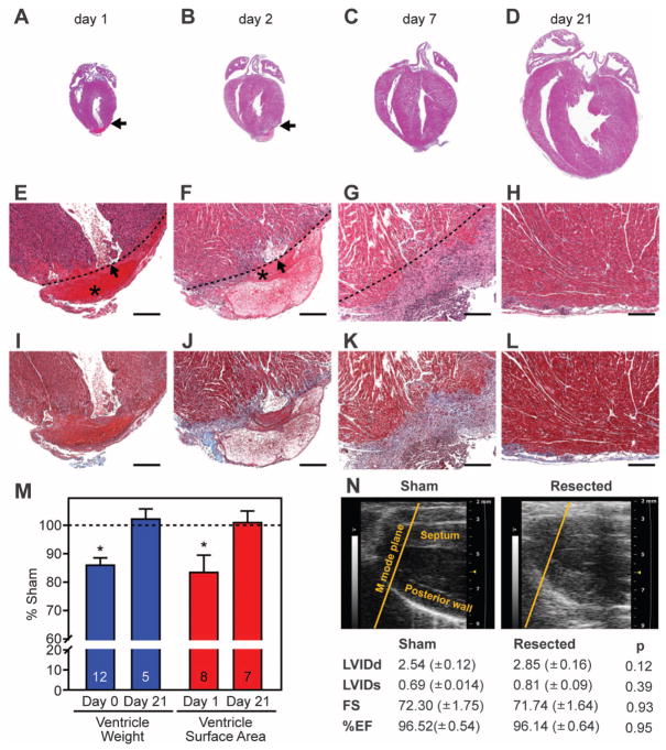
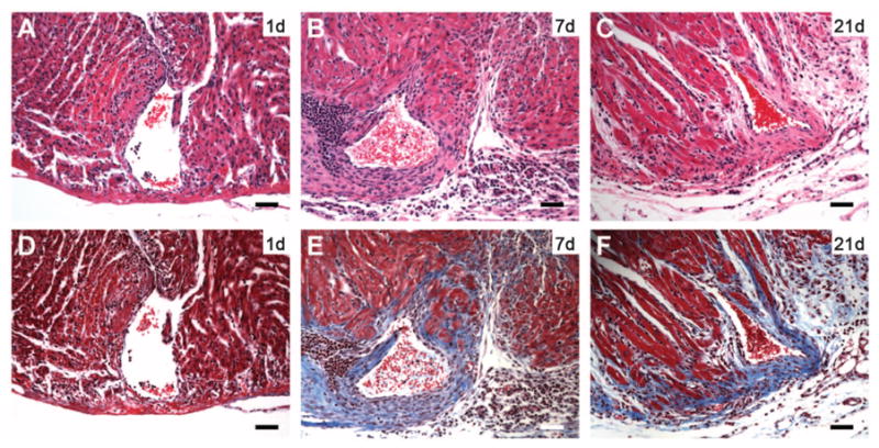
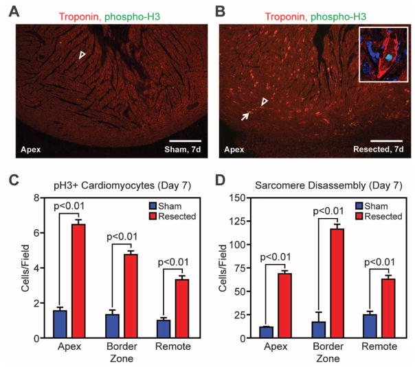
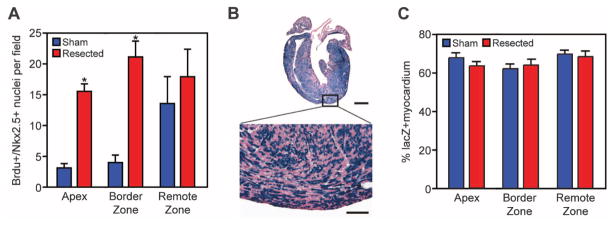
Certain fish and amphibians retain a robust capacity for cardiac regeneration throughout life, but the same is not true of the adult mammalian heart. Whether the capacity for cardiac regeneration is absent in mammals or whether it exists and is switched off early after birth has been unclear. We found that the hearts of 1-day-old neonatal mice can regenerate after partial surgical resection, but this capacity is lost by 7 days of age. This regenerative response in 1-day-old mice was characterized by cardiomyocyte proliferation with minimal hypertrophy or fibrosis, thereby distinguishing it from repair processes. Genetic fate mapping indicated that the majority of cardiomyocytes within the regenerated tissue originated from preexisting cardiomyocytes. Echocardiography performed 2 months after surgery revealed that the regenerated ventricular apex had normal systolic function. Thus, for a brief period after birth, the mammalian heart appears to have the capacity to regenerate.

摘要

某些鱼类和两栖类动物在整个生命周期中都保持着强大的心脏再生能力,但成年哺乳动物的心脏并非如此。哺乳动物是否缺乏心脏再生能力,或者这种能力是否存在但在出生后早期就被关闭,一直不清楚。我们发现,1 天大的新生小鼠的心脏在部分外科切除后可以再生,但这种能力在 7 天大时就丧失了。1 天大的小鼠的这种再生反应表现为心肌细胞增殖,而很少有肥大或纤维化,从而将其与修复过程区分开来。遗传命运图谱表明,再生组织中的大多数心肌细胞都源自原有心肌细胞。心脏超声检查显示,手术后 2 个月,再生的心室顶部具有正常的收缩功能。因此,在出生后的短暂时间内,哺乳动物的心脏似乎具有再生能力。

https://cdn.ncbi.nlm.nih.gov/pmc/blobs/1b27/3099478/29f45cd5926a/nihms283139f4.jpg
https://cdn.ncbi.nlm.nih.gov/pmc/blobs/1b27/3099478/472012e72ca9/nihms283139f1.jpg
https://cdn.ncbi.nlm.nih.gov/pmc/blobs/1b27/3099478/52442b370f8d/nihms283139f2.jpg
https://cdn.ncbi.nlm.nih.gov/pmc/blobs/1b27/3099478/86b8f646d4d7/nihms283139f3.jpg
https://cdn.ncbi.nlm.nih.gov/pmc/blobs/1b27/3099478/29f45cd5926a/nihms283139f4.jpg

相似文献

1
Transient regenerative potential of the neonatal mouse heart.新生鼠心脏的短暂再生潜能。
Science. 2011 Feb 25;331(6020):1078-80. doi: 10.1126/science.1200708.
2
Early Regenerative Capacity in the Porcine Heart.猪心的早期再生能力。
Circulation. 2018 Dec 11;138(24):2798-2808. doi: 10.1161/CIRCULATIONAHA.117.031542.
3
Extending the time window of mammalian heart regeneration by thymosin beta 4.胸腺素β4延长哺乳动物心脏再生的时间窗口。
J Cell Mol Med. 2014 Dec;18(12):2417-24. doi: 10.1111/jcmm.12421. Epub 2014 Oct 6.
4
Prolonged Myocardial Regenerative Capacity in Neonatal Opossum.幼年负鼠的心肌再生能力延长。
Circulation. 2022 Jul 12;146(2):125-139. doi: 10.1161/CIRCULATIONAHA.121.055269. Epub 2022 May 26.
5
Neonatal Apex Resection Triggers Cardiomyocyte Proliferation, Neovascularization and Functional Recovery Despite Local Fibrosis.新生儿心尖切除术可触发心肌细胞增殖、新生血管形成和功能恢复,尽管存在局部纤维化。
Stem Cell Reports. 2018 Mar 13;10(3):860-874. doi: 10.1016/j.stemcr.2018.01.042. Epub 2018 Mar 1.
6
A systematic analysis of neonatal mouse heart regeneration after apical resection.对新生小鼠心尖切除术后心脏再生的系统分析。
J Mol Cell Cardiol. 2015 Feb;79:315-8. doi: 10.1016/j.yjmcc.2014.12.011. Epub 2014 Dec 19.
7
Persistent scarring and dilated cardiomyopathy suggest incomplete regeneration of the apex resected neonatal mouse myocardium--A 180 days follow up study.持续性瘢痕形成和扩张型心肌病提示新生小鼠心肌切除术后心尖部再生不完全——一项180天随访研究。
J Mol Cell Cardiol. 2016 Jan;90:47-52. doi: 10.1016/j.yjmcc.2015.11.031. Epub 2015 Nov 30.
8
Regenerative Potential of Neonatal Porcine Hearts.新生猪心脏的再生潜力。
Circulation. 2018 Dec 11;138(24):2809-2816. doi: 10.1161/CIRCULATIONAHA.118.034886.
9
Heart regeneration in zebrafish.斑马鱼的心脏再生
Science. 2002 Dec 13;298(5601):2188-90. doi: 10.1126/science.1077857.
10
The type of injury dictates the mode of repair in neonatal and adult heart.损伤类型决定了新生儿和成人心脏的修复方式。
J Am Heart Assoc. 2015 Jan 27;4(1):e001320. doi: 10.1161/JAHA.114.001320.

引用本文的文献

1
Cardiac lymphatics retain LYVE-1-dependent macrophages during neonatal mouse heart regeneration.在新生小鼠心脏再生过程中,心脏淋巴管保留了依赖于LYVE-1的巨噬细胞。
Nat Cardiovasc Res. 2025 Sep 17. doi: 10.1038/s44161-025-00711-4.
2
Maintaining the LYVE1 line through macrophage and lymphatic interplay in the regenerating neonatal heart.通过巨噬细胞与淋巴管在新生心脏再生中的相互作用维持LYVE1谱系。
Nat Cardiovasc Res. 2025 Sep 17. doi: 10.1038/s44161-025-00707-0.
3
The emerging role of tissue regulatory T cells in tissue repair and regeneration.

本文引用的文献

1
Zebrafish heart regeneration occurs by cardiomyocyte dedifferentiation and proliferation.斑马鱼的心脏再生是通过心肌细胞去分化和增殖来实现的。
Nature. 2010 Mar 25;464(7288):606-9. doi: 10.1038/nature08899.
2
Primary contribution to zebrafish heart regeneration by gata4(+) cardiomyocytes.Gata4(+) 心肌细胞对斑马鱼心脏再生的主要贡献。
Nature. 2010 Mar 25;464(7288):601-5. doi: 10.1038/nature08804.
3
Cardiomyocyte cell cycle control and growth estimation in vivo--an analysis based on cardiomyocyte nuclei.心肌细胞周期控制和体内生长估计——基于心肌细胞核的分析。
组织调节性T细胞在组织修复和再生中的新作用。
Front Immunol. 2025 Aug 29;16:1640113. doi: 10.3389/fimmu.2025.1640113. eCollection 2025.
4
Transcriptional regulation in heart development, disease and regeneration: reassessing the fetal gene hypothesis.心脏发育、疾病与再生中的转录调控:重新审视胎儿基因假说。
Nat Rev Cardiol. 2025 Sep 5. doi: 10.1038/s41569-025-01205-3.
5
Translational Control in Cardiac Pathophysiology and Therapeutic Development: When mRNA Meets the Heart.心脏病理生理学与治疗学发展中的翻译控制:当信使核糖核酸与心脏相遇时。
Int J Mol Sci. 2025 Aug 14;26(16):7863. doi: 10.3390/ijms26167863.
6
Connexin 43 suppression enhances contractile force in human iPSC-derived cardiac tissues.连接蛋白43的抑制增强了人诱导多能干细胞衍生心肌组织的收缩力。
Front Bioeng Biotechnol. 2025 Aug 8;13:1615953. doi: 10.3389/fbioe.2025.1615953. eCollection 2025.
7
LncRNA HSCHARME is altered in human cardiomyopathies and promotes stem cell-derived cardiomyogenesis via splicing regulation.长链非编码RNA HSCHARME在人类心肌病中发生改变,并通过剪接调控促进干细胞来源的心肌生成。
Nat Commun. 2025 Aug 23;16(1):7880. doi: 10.1038/s41467-025-62754-2.
8
Sirt4 Deficiency Promotes Cardiomyocyte Proliferation and Cardiac Repair.沉默调节蛋白4缺乏促进心肌细胞增殖和心脏修复。
J Cell Mol Med. 2025 Aug;29(16):e70741. doi: 10.1111/jcmm.70741.
9
Cardiovascular diseases in the elderly: possibilities for modulating autophagy using non-coding RNAs.老年人的心血管疾病:利用非编码RNA调节自噬的可能性
Front Cell Dev Biol. 2025 Jul 31;13:1520850. doi: 10.3389/fcell.2025.1520850. eCollection 2025.
10
Advances and Prospects in Understanding Vertebrate Cardiac Conduction System, Pacemaker Cell, and Cardiac Muscle Development: Toward Novel Biological Therapies.脊椎动物心脏传导系统、起搏细胞和心肌发育的研究进展与展望:迈向新型生物疗法
Muscles. 2023 Oct 12;2(4):338-352. doi: 10.3390/muscles2040026.
Cardiovasc Res. 2010 Jun 1;86(3):365-73. doi: 10.1093/cvr/cvq005. Epub 2010 Jan 13.
4
Neuregulin1/ErbB4 signaling induces cardiomyocyte proliferation and repair of heart injury.神经调节蛋白1/表皮生长因子受体4信号通路可诱导心肌细胞增殖并修复心脏损伤。
Cell. 2009 Jul 23;138(2):257-70. doi: 10.1016/j.cell.2009.04.060.
5
Evidence for cardiomyocyte renewal in humans.人类心肌细胞更新的证据。
Science. 2009 Apr 3;324(5923):98-102. doi: 10.1126/science.1164680.
6
Compensatory growth of healthy cardiac cells in the presence of diseased cells restores tissue homeostasis during heart development.在患病细胞存在的情况下,健康心脏细胞的代偿性生长可在心脏发育过程中恢复组织稳态。
Dev Cell. 2008 Oct;15(4):521-33. doi: 10.1016/j.devcel.2008.09.005.
7
Evidence from a genetic fate-mapping study that stem cells refresh adult mammalian cardiomyocytes after injury.一项基因命运图谱研究的证据表明,干细胞在损伤后可更新成年哺乳动物的心肌细胞。
Nat Med. 2007 Aug;13(8):970-4. doi: 10.1038/nm1618. Epub 2007 Jul 29.
8
Cardiac myocyte cell cycle control in development, disease, and regeneration.发育、疾病和再生过程中心肌细胞的细胞周期调控
Physiol Rev. 2007 Apr;87(2):521-44. doi: 10.1152/physrev.00032.2006.
9
Getting to the heart of regeneration in zebrafish.深入了解斑马鱼的再生核心。
Semin Cell Dev Biol. 2007 Feb;18(1):36-45. doi: 10.1016/j.semcdb.2006.11.009. Epub 2006 Nov 24.
10
A dynamic epicardial injury response supports progenitor cell activity during zebrafish heart regeneration.动态的心外膜损伤反应在斑马鱼心脏再生过程中支持祖细胞活性。
Cell. 2006 Nov 3;127(3):607-19. doi: 10.1016/j.cell.2006.08.052.