Department of Histology and Embryology, Jessenius Faculty of Medicine Martin, Comenius University, Martin, Slovak Republic.
Med Sci Monit. 2011 Feb 25;17(3):BR74-80. doi: 10.12659/msm.881442.
The tumor suppressor gene p53 is a key regulator of cell division and/or apoptosis. Survivin is a multifunctional member of the inhibitor of apoptosis family. Survivin and p53 represent diametrically opposed signals that influence the apoptotic pathway.
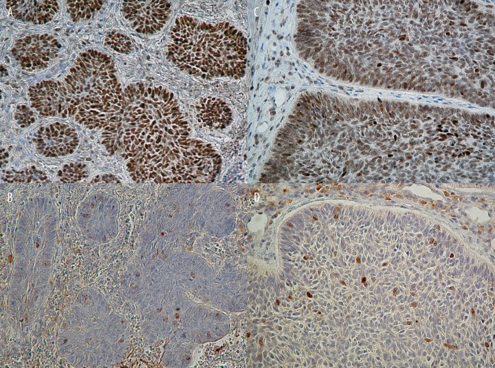
MATERIAL/METHODS: To determine the role of p53 and survivin in basal cell carcinoma (BCC), we evaluated the expression pattern of both proteins with regard to the percentage of positively immunostained tumor cells, the intensity of staining, and subcellular localization among 31 subjects with BCC.
Overexpression of p53 protein was found in 28 of 31 cases (90.3%), whereas survivin accumulation was seen in 27 (87.1%). For p53, moderate and/or strong immunoreactivity was seen in 20 of 28 cases (71.4%), and 26 of 28 cases (92.9%) showed more than 25% reactive tumor cells. Nuclear p53 staining was detected in 23 of 28 cases (82.1%), whereas combined nuclear and cytoplasmic localization was found in only 5 of 28 cases (17.9%). Survivin revealed mild intensity of immunoreaction in 22 of 27 cases (71%), and 25 of 27 cases (92.6%) showed less than 25% labeled tumor cells. Combined nuclear and cytoplasmic survivin localization was present in 26 of 27 cases (96.3%). Statistically significant differences were detected in the assessed expression parameters between those proteins.
Our results suggest that overexpression of wild type p53 protein may suppress the expression of survivin and its antiapoptotic activity in BCC cells.
抑癌基因 p53 是细胞分裂和/或凋亡的关键调节因子。生存素是凋亡抑制因子家族的多功能成员。生存素和 p53 代表了影响凋亡途径的截然相反的信号。
材料/方法:为了确定 p53 和生存素在基底细胞癌 (BCC) 中的作用,我们评估了这两种蛋白质的表达模式,包括阳性免疫染色肿瘤细胞的百分比、染色强度和亚细胞定位,共涉及 31 例 BCC 患者。
p53 蛋白过表达见于 31 例中的 28 例 (90.3%),而生存素积累见于 27 例 (87.1%)。p53 的中度和/或强免疫反应性见于 28 例中的 20 例 (71.4%),28 例中有 26 例 (92.9%)显示超过 25%的反应性肿瘤细胞。23 例中的 28 例 (82.1%)检测到核 p53 染色,而只有 28 例中的 5 例 (17.9%)发现核质共定位。27 例中的 22 例 (71%)显示生存素免疫反应性轻度,27 例中有 25 例 (92.6%)显示小于 25%的标记肿瘤细胞。27 例中的 26 例 (96.3%)存在核质共定位的生存素。这两种蛋白质的评估表达参数之间存在统计学显著差异。
我们的结果表明,野生型 p53 蛋白的过表达可能抑制 BCC 细胞中生存素的表达及其抗凋亡活性。