Department of Prosthodontics, Malmo University, Malmo, Sweden.
BMC Oral Health. 2011 Mar 8;11:8. doi: 10.1186/1472-6831-11-8.
The soft tissue around dental implants forms a barrier between the oral environment and the peri-implant bone and a crucial factor for long-term success of therapy is development of a good abutment/soft-tissue seal. Sol-gel derived nanoporous TiO2 coatings have been shown to enhance soft-tissue attachment but their effect on adhesion and biofilm formation by oral bacteria is unknown.
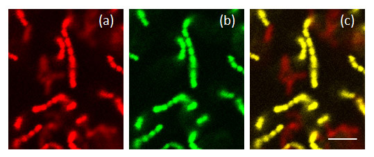
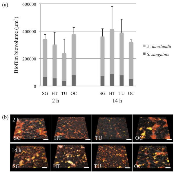
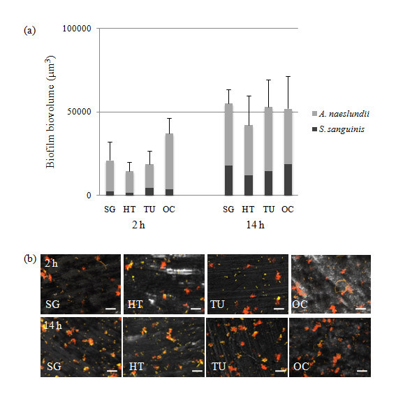
We have investigated how the properties of surfaces that may be used on abutments: turned titanium, sol-gel nanoporous TiO2 coated surfaces and anodized Ca2+ modified surfaces, affect biofilm formation by two early colonizers of the oral cavity: Streptococcus sanguinis and Actinomyces naeslundii. The bacteria were detected using 16S rRNA fluorescence in situ hybridization together with confocal laser scanning microscopy.
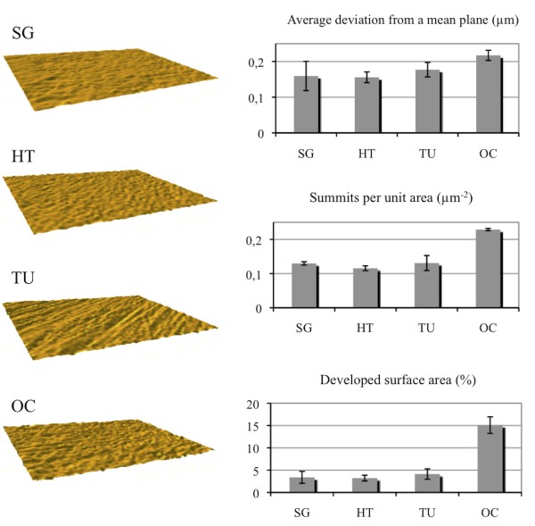
Interferometry and atomic force microscopy revealed all the surfaces to be smooth (Sa≤0.22 μm). Incubation with a consortium of S. sanguinis and A. naeslundii showed no differences in adhesion between the surfaces over 2 hours. After 14 hours, the level of biofilm growth was low and again, no differences between the surfaces were seen. The presence of saliva increased the biofilm biovolume of S. sanguinis and A. naeslundii ten-fold compared to when saliva was absent and this was due to increased adhesion rather than biofilm growth.
Nano-topographical modification of smooth titanium surfaces had no effect on adhesion or early biofilm formation by S. sanguinis and A. naeslundii as compared to turned surfaces or those treated with anodic oxidation in the presence of Ca2+. The presence of saliva led to a significantly greater biofilm biovolume but no significant differences were seen between the test surfaces. These data thus suggest that modification with sol-gel derived nanoporous TiO2, which has been shown to improve osseointegration and soft-tissue healing in vivo, does not cause greater biofilm formation by the two oral commensal species tested than the other surfaces.
种植体周围的软组织在口腔环境和种植体周围骨之间形成了一道屏障,而治疗长期成功的一个关键因素是形成良好的基台/软组织密封。溶胶-凝胶衍生的纳米多孔 TiO2 涂层已被证明可以增强软组织附着,但它们对口腔细菌粘附和生物膜形成的影响尚不清楚。
我们研究了可能用于基台的表面特性:车削钛、溶胶-凝胶纳米多孔 TiO2 涂层表面和 Ca2+ 改性的阳极氧化表面如何影响口腔早期定植者两种细菌:血链球菌和内氏放线菌的生物膜形成。使用 16S rRNA 荧光原位杂交结合共焦激光扫描显微镜检测细菌。
干涉测量和原子力显微镜显示所有表面均光滑(Sa≤0.22μm)。用血链球菌和内氏放线菌的混合物孵育 2 小时后,各表面间的粘附无差异。14 小时后,生物膜生长水平较低,各表面之间仍未见差异。与无唾液相比,唾液的存在使血链球菌和内氏放线菌的生物膜生物量增加了十倍,这是由于粘附增加而不是生物膜生长。
与车削表面或在 Ca2+ 存在下进行阳极氧化处理的表面相比,光滑钛表面的纳米形貌修饰对血链球菌和内氏放线菌的粘附或早期生物膜形成没有影响。唾液的存在导致生物膜生物量显著增加,但在测试表面之间未观察到显著差异。因此,这些数据表明,溶胶-凝胶衍生的纳米多孔 TiO2 改性虽然已被证明可改善体内骨整合和软组织愈合,但不会导致比其他表面形成更多的两种口腔共生菌的生物膜。