• 文献检索
  • 文档翻译
  • 深度研究
  • 学术资讯
  • Suppr Zotero 插件Zotero 插件
  • 邀请有礼
  • 套餐&价格
  • 历史记录
应用&插件
Suppr Zotero 插件Zotero 插件浏览器插件Mac 客户端Windows 客户端微信小程序
定价
高级版会员购买积分包购买API积分包
服务
文献检索文档翻译深度研究API 文档MCP 服务
关于我们
关于 Suppr公司介绍联系我们用户协议隐私条款
关注我们

Suppr 超能文献

核心技术专利:CN118964589B侵权必究
粤ICP备2023148730 号-1Suppr @ 2026

文献检索

告别复杂PubMed语法,用中文像聊天一样搜索,搜遍4000万医学文献。AI智能推荐,让科研检索更轻松。

立即免费搜索

文件翻译

保留排版,准确专业,支持PDF/Word/PPT等文件格式,支持 12+语言互译。

免费翻译文档

深度研究

AI帮你快速写综述,25分钟生成高质量综述,智能提取关键信息,辅助科研写作。

立即免费体验

基于受控制的 3D 纳米结构介导的表面增强拉曼散射的细胞内状态分析。

Analysis of intracellular state based on controlled 3D nanostructures mediated surface enhanced Raman scattering.

机构信息

Interdisciplinary Program of Integrated Biotechnology, Sogang University, Seoul, Republic of Korea.

出版信息

PLoS One. 2011 Feb 24;6(2):e15836. doi: 10.1371/journal.pone.0015836.

DOI:10.1371/journal.pone.0015836
PMID:21390213
原文链接:https://pmc.ncbi.nlm.nih.gov/articles/PMC3044723/
Abstract

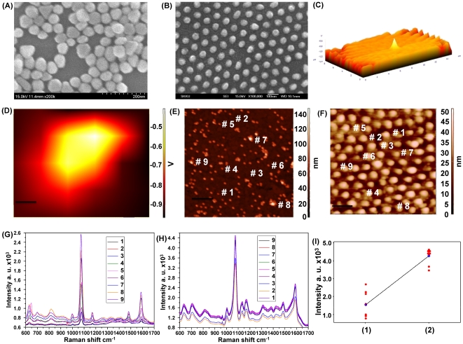
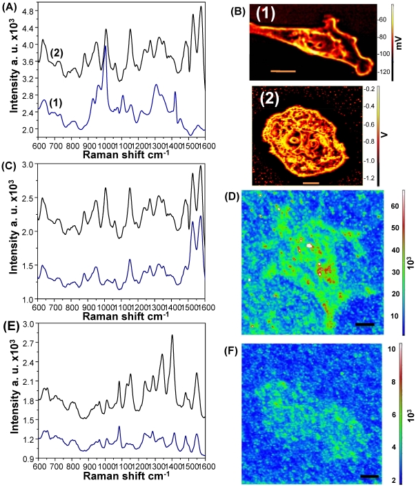
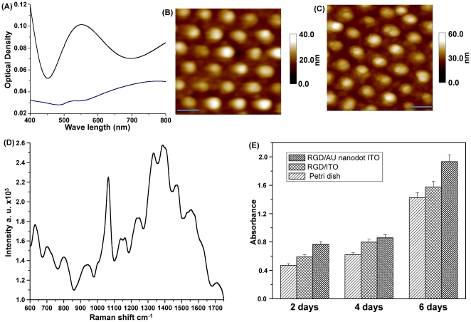
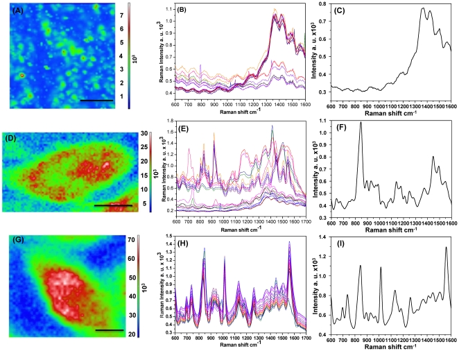
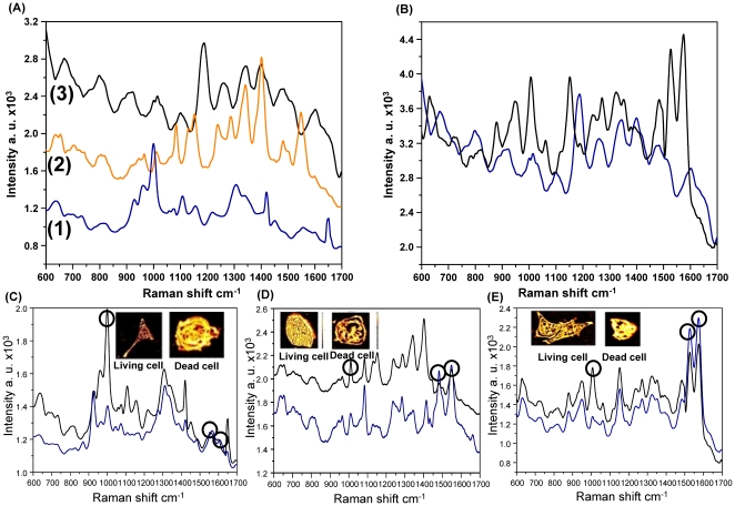
Near-infrared surface-enhanced Raman spectroscopy (SERS) is a powerful technique for analyzing the chemical composition within a single living cell at unprecedented resolution. However, current SERS methods employing uncontrollable colloidal metal particles or non-uniformly distributed metal particles on a substrate as SERS-active sites show relatively low reliability and reproducibility. Here, we report a highly-ordered SERS-active surface that is provided by a gold nano-dots array based on thermal evaporation of gold onto an ITO surface through a nanoporous alumina mask. This new combined technique showed a broader distribution of hot spots and a higher signal-to-noise ratio than current SERS techniques due to the highly reproducible and uniform geometrical structures over a large area. This SERS-active surface was applied as cell culture system to study living cells in situ within their culture environment without any external preparation processes. We applied this newly developed method to cell-based research to differentiate cell lines, cells at different cell cycle stages, and live/dead cells. The enhanced Raman signals achieved from each cell, which represent the changes in biochemical compositions, enabled differentiation of each state and the conditions of the cells. This SERS technique employing a tightly controlled nanostructure array can potentially be applied to single cell analysis, early cancer diagnosis and cell physiology research.

摘要

近红外表面增强拉曼光谱(SERS)是一种强大的技术,可以以前所未有的分辨率分析单个活细胞内的化学成分。然而,目前采用不可控胶体金属颗粒或在基底上不均匀分布的金属颗粒作为 SERS 活性位点的 SERS 方法,显示出相对较低的可靠性和重现性。在这里,我们报告了一种高度有序的 SERS 活性表面,它是通过在 ITO 表面上通过纳米多孔氧化铝掩模热蒸发金而提供的基于金纳米点阵列的。由于在大面积上具有高度可重复且均匀的几何结构,这种新的组合技术显示出更广泛的热点分布和更高的信噪比,优于当前的 SERS 技术。这种 SERS 活性表面被用作细胞培养系统,用于在其培养环境中对活细胞进行原位研究,而无需任何外部制备过程。我们将这种新开发的方法应用于基于细胞的研究,以区分细胞系、不同细胞周期阶段的细胞以及活/死细胞。从每个细胞获得的增强拉曼信号代表了生化成分的变化,从而能够区分每个状态和细胞的条件。这种采用紧密控制的纳米结构阵列的 SERS 技术可能适用于单细胞分析、早期癌症诊断和细胞生理学研究。

https://cdn.ncbi.nlm.nih.gov/pmc/blobs/d7e6/3044723/f71d18082630/pone.0015836.g005.jpg
https://cdn.ncbi.nlm.nih.gov/pmc/blobs/d7e6/3044723/d132a8d06f5c/pone.0015836.g001.jpg
https://cdn.ncbi.nlm.nih.gov/pmc/blobs/d7e6/3044723/2575b1958506/pone.0015836.g002.jpg
https://cdn.ncbi.nlm.nih.gov/pmc/blobs/d7e6/3044723/3469253ccfca/pone.0015836.g003.jpg
https://cdn.ncbi.nlm.nih.gov/pmc/blobs/d7e6/3044723/ec89f0e4caf0/pone.0015836.g004.jpg
https://cdn.ncbi.nlm.nih.gov/pmc/blobs/d7e6/3044723/f71d18082630/pone.0015836.g005.jpg

相似文献

1
Analysis of intracellular state based on controlled 3D nanostructures mediated surface enhanced Raman scattering.基于受控制的 3D 纳米结构介导的表面增强拉曼散射的细胞内状态分析。
PLoS One. 2011 Feb 24;6(2):e15836. doi: 10.1371/journal.pone.0015836.
2
Developing Hollow-Channel Gold Nanoflowers as Trimodal Intracellular Nanoprobes.研制中空通道金纳米花作为三模态细胞内纳米探针。
Int J Mol Sci. 2018 Aug 8;19(8):2327. doi: 10.3390/ijms19082327.
3
Hot spots in different metal nanostructures for plasmon-enhanced Raman spectroscopy.不同金属纳米结构中的热点用于等离子体增强拉曼光谱学。
Nanoscale. 2013 Nov 21;5(22):10794-805. doi: 10.1039/c3nr02924g. Epub 2013 Oct 11.
4
Nanoarchitecture Based SERS for Biomolecular Fingerprinting and Label-Free Disease Markers Diagnosis.基于纳米结构的 SERS 用于生物分子指纹图谱和无标记疾病标志物诊断。
Acc Chem Res. 2016 Dec 20;49(12):2725-2735. doi: 10.1021/acs.accounts.6b00384. Epub 2016 Dec 8.
5
In situ fabrication of 3D Ag@ZnO nanostructures for microfluidic surface-enhanced Raman scattering systems.用于微流控表面增强拉曼散射系统的3D Ag@ZnO纳米结构的原位制备。
ACS Nano. 2014 Dec 23;8(12):12175-84. doi: 10.1021/nn503826r. Epub 2014 Nov 17.
6
In situ intracellular spectroscopy with surface enhanced Raman spectroscopy (SERS)-enabled nanopipettes.基于表面增强拉曼光谱(SERS)的纳米电极管的细胞内原位光谱技术。
ACS Nano. 2009 Nov 24;3(11):3529-36. doi: 10.1021/nn9010768.
7
Large area flexible SERS active substrates using engineered nanostructures.采用工程化纳米结构的大面积柔性 SERS 活性衬底。
Nanoscale. 2011 Jul;3(7):2903-8. doi: 10.1039/c1nr10265f. Epub 2011 May 31.
8
Gold nanoaggregates for probing single-living cell based on surface-enhanced Raman spectroscopy.基于表面增强拉曼光谱的用于探测单个活细胞的金纳米聚集体
J Biomed Opt. 2015 May;20(5):051005. doi: 10.1117/1.JBO.20.5.051005.
9
Highly uniform and reproducible surface-enhanced Raman scattering from DNA-tailorable nanoparticles with 1-nm interior gap.具有 1nm 内间隙的可通过 DNA 修饰的纳米粒子实现高度均匀且可重现的表面增强拉曼散射。
Nat Nanotechnol. 2011 May 29;6(7):452-60. doi: 10.1038/nnano.2011.79.
10
3D aluminum/silver hierarchical nanostructure with large areas of dense hot spots for surface-enhanced raman scattering.具有大面积密集热点的 3D 铝/银分层纳米结构用于表面增强拉曼散射。
Electrophoresis. 2019 Dec;40(23-24):3123-3131. doi: 10.1002/elps.201900285. Epub 2019 Oct 14.

引用本文的文献

1
Dielectrophoresis-Based SERS Sensors for the Detection of Cancer Cells in Microfluidic Chips.基于介电泳的微流控芯片上用于检测癌细胞的 SERS 传感器。
Biosensors (Basel). 2022 Aug 25;12(9):681. doi: 10.3390/bios12090681.
2
Minimally invasive detection of cancer using metabolic changes in tumor-associated natural killer cells with Oncoimmune probes.使用 Oncoimmune 探针检测肿瘤相关自然杀伤细胞代谢变化进行微创癌症检测。
Nat Commun. 2022 Aug 4;13(1):4527. doi: 10.1038/s41467-022-32308-x.
3
Characterization of Labeled Gold Nanoparticles for Surface-Enhanced Raman Scattering.

本文引用的文献

1
Detection of effect of chemotherapeutic agents to cancer cells on gold nanoflower patterned substrate using surface-enhanced Raman scattering and cyclic voltammetry.利用表面增强拉曼散射和循环伏安法检测金纳米花图案化基底上化疗药物对癌细胞的作用。
Biosens Bioelectron. 2010 Dec 15;26(4):1486-92. doi: 10.1016/j.bios.2010.07.089. Epub 2010 Jul 30.
2
Effects of nanopatterned RGD peptide layer on electrochemical detection of neural cell chip.纳米图案化 RGD 肽层对神经细胞芯片电化学检测的影响。
Biosens Bioelectron. 2010 Dec 15;26(4):1359-65. doi: 10.1016/j.bios.2010.07.049. Epub 2010 Jul 21.
3
Nuclear targeting of gold nanoparticles in cancer cells induces DNA damage, causing cytokinesis arrest and apoptosis.
用于表面增强拉曼散射的标记金纳米粒子的特性描述。
Molecules. 2022 Jan 28;27(3):892. doi: 10.3390/molecules27030892.
4
Efficient Capture and Raman Analysis of Circulating Tumor Cells by Nano-Undulated AgNPs-rGO Composite SERS Substrates.纳米波纹状AgNPs-rGO复合表面增强拉曼散射基底对循环肿瘤细胞的高效捕获与拉曼分析
Sensors (Basel). 2020 Sep 7;20(18):5089. doi: 10.3390/s20185089.
5
Detection of circulating tumor cells in blood by shell-isolated nanoparticle - enhanced Raman spectroscopy (SHINERS) in microfluidic device.通过微流控装置中的壳层分离纳米颗粒增强拉曼光谱法(SHINERS)检测血液中的循环肿瘤细胞。
Sci Rep. 2019 Jun 25;9(1):9267. doi: 10.1038/s41598-019-45629-7.
6
Detection of Circulating Tumor Cells Using Membrane-Based SERS Platform: A New Diagnostic Approach for 'Liquid Biopsy'.基于膜的表面增强拉曼散射平台检测循环肿瘤细胞:一种“液体活检”的新诊断方法
Nanomaterials (Basel). 2019 Mar 5;9(3):366. doi: 10.3390/nano9030366.
7
Nanostructured surfaces for analysis of anticancer drug and cell diagnosis based on electrochemical and SERS tools.基于电化学和表面增强拉曼光谱工具的用于抗癌药物分析和细胞诊断的纳米结构表面
Nano Converg. 2018;5(1):11. doi: 10.1186/s40580-018-0143-4. Epub 2018 Apr 24.
8
Gold Nanoparticles in Single-Cell Analysis for Surface Enhanced Raman Scattering.用于表面增强拉曼散射的单细胞分析中的金纳米颗粒
Molecules. 2016 Nov 25;21(12):1617. doi: 10.3390/molecules21121617.
9
Beneficial effects of cerium oxide nanoparticles in development of chondrocyte-seeded hydrogel constructs and cellular response to interleukin insults.氧化铈纳米颗粒在软骨细胞接种水凝胶构建物发育及细胞对白介素刺激的反应中的有益作用。
Tissue Eng Part A. 2014 Nov;20(21-22):2908-19. doi: 10.1089/ten.TEA.2013.0592. Epub 2014 Jun 25.
10
A simple and rapid detection of tissue adhesive-induced biochemical changes in cells and DNA using Raman spectroscopy.利用拉曼光谱法简单快速地检测组织黏合剂诱导的细胞和DNA中的生化变化。
Biomed Opt Express. 2013 Oct 29;4(11):2673-82. doi: 10.1364/BOE.4.002673. eCollection 2013.
金纳米颗粒在癌细胞中的核靶向导致 DNA 损伤,引起细胞分裂停滞和细胞凋亡。
J Am Chem Soc. 2010 Feb 10;132(5):1517-9. doi: 10.1021/ja9102698.
4
Cell-based chip for the detection of anticancer effect on HeLa cells using cyclic voltammetry.基于细胞的芯片,用于通过循环伏安法检测对HeLa细胞的抗癌作用。
Biosens Bioelectron. 2009 Jan 1;24(5):1259-65. doi: 10.1016/j.bios.2008.07.037. Epub 2008 Jul 31.
5
Measurement of the distribution of site enhancements in surface-enhanced Raman scattering.表面增强拉曼散射中位点增强分布的测量。
Science. 2008 Jul 18;321(5887):388-92. doi: 10.1126/science.1159499. Epub 2008 Jun 26.
6
Clean substrates prepared by chemical adsorption of iodide followed by electrochemical oxidation for surface-enhanced Raman spectroscopic study of cell membrane.通过碘化物化学吸附,随后进行电化学氧化制备的清洁基底,用于细胞膜的表面增强拉曼光谱研究。
Anal Chem. 2008 Jul 1;80(13):5118-25. doi: 10.1021/ac8003083. Epub 2008 May 20.
7
Diagnostic potential of near-infrared Raman spectroscopy in the stomach: differentiating dysplasia from normal tissue.近红外拉曼光谱在胃部的诊断潜力:区分发育异常组织与正常组织。
Br J Cancer. 2008 Jan 29;98(2):457-65. doi: 10.1038/sj.bjc.6604176. Epub 2008 Jan 15.
8
Ordered metal nanohole arrays made by a two-step replication of honeycomb structures of anodic alumina.两步复制法制备有序金属纳米孔阵列及其模板氧化铝的蜂窝结构
Science. 1995 Jun 9;268(5216):1466-8. doi: 10.1126/science.268.5216.1466.
9
Cancer cells assemble and align gold nanorods conjugated to antibodies to produce highly enhanced, sharp, and polarized surface Raman spectra: a potential cancer diagnostic marker.癌细胞组装并排列与抗体结合的金纳米棒,以产生高度增强、尖锐且偏振的表面拉曼光谱:一种潜在的癌症诊断标志物。
Nano Lett. 2007 Jun;7(6):1591-7. doi: 10.1021/nl070472c. Epub 2007 May 3.
10
Metabolic fingerprinting in disease diagnosis: biomedical applications of infrared and Raman spectroscopy.疾病诊断中的代谢指纹图谱:红外光谱和拉曼光谱的生物医学应用
Analyst. 2006 Aug;131(8):875-85. doi: 10.1039/b602376m. Epub 2006 Apr 25.