Department of Neurology, Erasmus University Medical Center, Room H-639, 's-Gravendijkwal 230, 3015 CE Rotterdam, The Netherlands.
J Neurol. 2011 Aug;258(8):1507-12. doi: 10.1007/s00415-011-5970-8. Epub 2011 Mar 12.
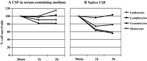
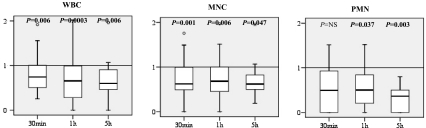
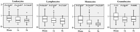
Immediately after sampling, leukocyte counts in native cerebrospinal fluid (CSF) start to decrease rapidly. As the time lapse between CSF collection to analysis is not routinely registered, the clinical significance of decreasing cell counts in native CSF is not known. Earlier data suggest that addition of serum-containing medium to CSF directly after sampling prevents this rapid decrease in leukocyte counts and, thus, may improve the accuracy of CSF cell counting and cell characterization. Here, we prospectively examined the effect of storage time after lumbar puncture on counts of leukocytes and their major subsets in both native CSF and after immediate addition of serum-containing medium, measured by flow cytometry and microscopy. We collected CSF samples of 69 patients in tubes with and tubes without serum-containing medium and determined counts of leukocytes and subsets at 30 minutes, 1 hour, and 5 hours after sampling. Compared to cell counts at 30 minutes, no significant decrease in cell number was observed in CSF with serum-containing medium 1 and 5 hours after sampling, except for the granulocytes at 1 hour. In native CSF, approximately 50% of leukocytes and all their subsets were lost after 1 hour, both in flow cytometric and microscopic counting. In 6/7 (86%) samples with mild pleocytosis (5-15 × 10(6) leukocytes/l), native CSF at 1 hour was incorrectly diagnosed as normocellular. In conclusion, addition of serum-containing medium to CSF directly after sampling prevents cell loss and allows longer preservation of CSF cells prior to analysis, both for microscopic and flow cytometric enumeration. We suggest that this protocol results in more accurate CSF cell counts and may prevent incorrect conclusions based on underestimated CSF cell counts.
采样后,原生脑脊髓液(CSF)中的白细胞计数会迅速开始下降。由于 CSF 采集到分析之间的时间间隔未被常规记录,因此,原生 CSF 中细胞计数下降的临床意义尚不清楚。早期数据表明,采样后直接向 CSF 中添加含血清的培养基可防止白细胞计数的快速下降,从而可能提高 CSF 细胞计数和细胞特征分析的准确性。在此,我们前瞻性地研究了腰椎穿刺后储存时间对原生 CSF 和立即添加含血清培养基后 CSF 中白细胞及其主要亚群计数的影响,通过流式细胞术和显微镜进行了测量。我们在含血清和不含血清的管中收集了 69 例患者的 CSF 样本,并在采样后 30 分钟、1 小时和 5 小时测定白细胞和亚群的计数。与 30 分钟时的细胞计数相比,除 1 小时时的粒细胞外,添加含血清培养基后 1 和 5 小时 CSF 中的细胞数没有明显下降。在原生 CSF 中,除 1 小时外,大约 50%的白细胞及其所有亚群在 1 小时后丢失,无论是在流式细胞术计数还是显微镜计数中。在 6/7(86%)轻度白细胞增多(5-15×10(6)个白细胞/L)的样本中,1 小时时的原生 CSF 被错误地诊断为正常细胞。总之,采样后直接向 CSF 中添加含血清的培养基可防止细胞丢失,并允许在分析之前更长时间地保存 CSF 细胞,无论是用于显微镜还是流式细胞术计数。我们建议,该方案可得到更准确的 CSF 细胞计数,并可防止基于低估的 CSF 细胞计数得出错误的结论。