Department of Physiology, Faculty of Medicine and Health Sciences, University of Auckland, Auckland, New Zealand.
PLoS One. 2011 Mar 9;6(3):e17901. doi: 10.1371/journal.pone.0017901.
The cardiac myocyte t-tubular system ensures rapid, uniform cell activation and several experimental lines of evidence suggest changes in the t-tubular system and associated excitation-contraction coupling proteins may occur in heart failure.
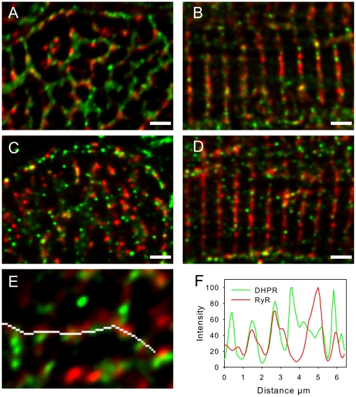
The organization of t-tubules, L-type calcium channels (DHPRs), ryanodine receptors (RyRs) and contractile machinery were examined in fixed ventricular tissue samples from both normal and failing hearts (idiopathic (non-ischemic) dilated cardiomyopathy) using high resolution fluorescent imaging. Wheat germ agglutinin (WGA), Na-Ca exchanger, DHPR and caveolin-3 labels revealed a shift from a predominantly transverse orientation to oblique and axial directions in failing myocytes. In failure, dilation of peripheral t-tubules occurred and a change in the extent of protein glycosylation was evident. There was no change in the fractional area occupied by myofilaments (labeled with phalloidin) but there was a small reduction in the number of RyR clusters per unit area. The general relationship between DHPRs and RyR was not changed and RyR labeling overlapped with 51±3% of DHPR labeling in normal hearts. In longitudinal (but not transverse) sections there was an ∼30% reduction in the degree of colocalization between DHPRs and RyRs as measured by Pearson's correlation coefficient in failing hearts.
The results show that extensive remodelling of the t-tubular network and associated excitation-contraction coupling proteins occurs in failing human heart. These changes may contribute to abnormal calcium handling in heart failure. The general organization of the t-system and changes observed in failure samples have subtle differences to some animal models although the general direction of changes are generally similar.
心肌细胞 T 管系统确保了快速、均匀的细胞激活,并且有几项实验证据表明,T 管系统和相关的兴奋-收缩偶联蛋白可能会发生变化。
使用高分辨率荧光成像技术,在正常和衰竭心脏(特发性(非缺血性)扩张型心肌病)的固定心室组织样本中检查 T 管、L 型钙通道(DHPRs)、ryanodine 受体(RyRs)和收缩机制的组织。小麦胚凝集素(WGA)、Na-Ca 交换器、DHPR 和 caveolin-3 标记显示,衰竭心肌细胞中的 T 管从主要的横向方向转变为斜向和轴向方向。在衰竭时,外周 T 管扩张,并且蛋白质糖基化程度发生变化。肌丝(用鬼笔环肽标记)占据的分数面积没有变化,但每个单位面积的 RyR 簇数量略有减少。DHPR 和 RyR 之间的一般关系没有改变,并且 RyR 标记与正常心脏中的 51±3%的 DHPR 标记重叠。在纵向(但不是横向)部分中,衰竭心脏中 DHPR 和 RyR 之间的共定位程度(通过 Pearson 相关系数测量)降低了约 30%。
结果表明,在衰竭的人心肌中,T 管网络和相关的兴奋-收缩偶联蛋白发生了广泛的重塑。这些变化可能导致心力衰竭时钙处理异常。尽管变化的总体方向通常相似,但 T 系统的一般组织和在衰竭样本中观察到的变化与一些动物模型略有不同。