Bowles Center for Alcohol Studies, University of North Carolina at Chapel Hill, Chapel Hill, NC 27599, USA.
Neuropsychol Rev. 2011 Jun;21(2):167-85. doi: 10.1007/s11065-011-9164-z. Epub 2011 Mar 29.
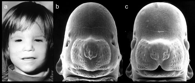
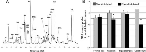
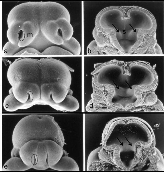
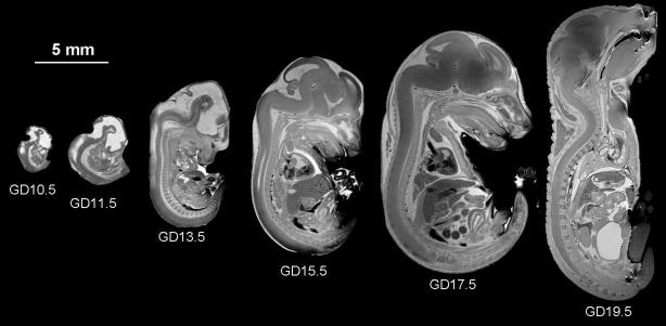
Magnetic resonance imaging (MRI) techniques, such as magnetic resonance microscopy (MRM), diffusion tensor imaging (DTI), and magnetic resonance spectroscopy (MRS), have recently been applied to the study of both normal and abnormal structure and neurochemistry in small animals. Herein, findings from studies in which these methods have been used for the examination of animal models of Fetal Alcohol Spectrum Disorder (FASD) are discussed. Emphasis is placed on results of imaging studies in fetal and postnatal mice that have highlighted the developmental stage dependency of prenatal ethanol exposure-induced CNS defects. Consideration is also given to the promise of methodological advances to allow in vivo studies of aberrant brain and behavior relationships in model animals and to the translational nature of this work.
磁共振成像(MRI)技术,如磁共振显微镜(MRM)、扩散张量成像(DTI)和磁共振波谱(MRS),最近已应用于小动物正常和异常结构及神经化学的研究。本文讨论了这些方法在胎儿酒精谱系障碍(FASD)动物模型检查中应用的研究结果。重点介绍了在胎儿和新生小鼠成像研究中的结果,这些结果强调了产前乙醇暴露引起的中枢神经系统缺陷与发育阶段的相关性。同时还考虑了方法学进展的前景,以允许在模型动物中进行异常大脑和行为关系的体内研究,以及这项工作的转化性质。