Laboratory of Pharmacology, Department of Oncology, Biology and Genetics University of Genova, Genova, Italy.
Cell Death Dis. 2011 Mar 31;2(3):e138. doi: 10.1038/cddis.2011.21.
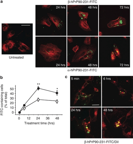
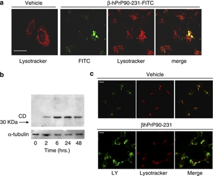
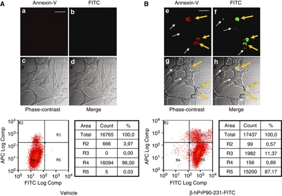
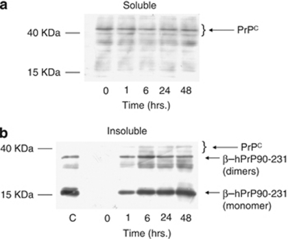
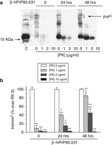
To define the mechanisms by which hPrP90-231 induces cell death, we analyzed its interaction with living cells and monitored its intracellular fate. Treatment of SH-SY5Y cells with fluorescein-5-isothiocyanate (FITC)-conjugated hPrP90-231 caused the accumulation of cytosolic aggregates of the prion protein fragment that increased in number and size in a time-dependent manner. The formation of large intracellular hPrP90-231 aggregates correlated with the activation of apoptosis. hPrP90-231 aggregates occurred within lysotracker-positive vesicles and induced the formation of activated cathepsin D (CD), indicating that hPrP90-231 is partitioned into the endosomal-lysosomal system structures, activating the proteolytic machinery. Remarkably, the inhibition of CD activity significantly reduced hPrP-90-231-dependent apoptosis. Internalized hPrP90-231 forms detergent-insoluble and SDS-stable aggregates, displaying partial resistance to proteolysis. By confocal microscopy analysis of lucifer yellow (LY) intracellular partition, we show that hPrP90-231 accumulation induces lysosome destabilization and loss of lysosomal membrane impermeability. In fact, although control cells evidenced a vesicular pattern of LY fluorescence (index of healthy lysosomes), hPrP90-231-treated cells showed diffuse cytosolic fluorescence, indicating LY diffusion through damaged lysosomes. In conclusion, these data indicate that exogenously added hPrP90-231 forms intralysosomal deposits having features of insoluble, protease-resistant aggregates and could trigger a lysosome-mediated apoptosis by inducing lysosome membrane permeabilization, followed by the release of hydrolytic enzymes.
为了明确朊蛋白 90-231 诱导细胞死亡的机制,我们分析了它与活细胞的相互作用,并监测了其细胞内命运。用异硫氰酸荧光素(FITC)标记的朊蛋白 90-231 处理 SH-SY5Y 细胞,导致朊蛋白片段的细胞质聚集物的积累,这些聚集物的数量和大小随时间呈依赖性增加。大的细胞内朊蛋白 90-231 聚集物的形成与细胞凋亡的激活相关。朊蛋白 90-231 聚集物发生在溶酶体阳性小泡内,并诱导激活的组织蛋白酶 D(CD)的形成,表明朊蛋白 90-231 被分配到内体-溶酶体系统结构中,激活了蛋白水解机制。值得注意的是,CD 活性的抑制显著减少了依赖于朊蛋白 90-231 的细胞凋亡。内化的朊蛋白 90-231 形成去污剂不溶性和 SDS 稳定的聚集物,显示出部分抵抗蛋白水解的能力。通过对荧光素黄(LY)细胞内分布的共焦显微镜分析,我们表明朊蛋白 90-231 的积累诱导溶酶体不稳定和溶酶体膜通透性丧失。事实上,尽管对照细胞显示出 LY 荧光的囊泡模式(健康溶酶体的指标),但朊蛋白 90-231 处理的细胞显示出弥漫的细胞质荧光,表明 LY 通过受损的溶酶体扩散。总之,这些数据表明,外加的朊蛋白 90-231 形成具有不溶性、抗蛋白酶聚集物特征的溶酶体内沉积物,并通过诱导溶酶体膜通透性,随后释放水解酶,可能引发溶酶体介导的细胞凋亡。