• 文献检索
  • 文档翻译
  • 深度研究
  • 学术资讯
  • Suppr Zotero 插件Zotero 插件
  • 邀请有礼
  • 套餐&价格
  • 历史记录
应用&插件
Suppr Zotero 插件Zotero 插件浏览器插件Mac 客户端Windows 客户端微信小程序
定价
高级版会员购买积分包购买API积分包
服务
文献检索文档翻译深度研究API 文档MCP 服务
关于我们
关于 Suppr公司介绍联系我们用户协议隐私条款
关注我们

Suppr 超能文献

核心技术专利:CN118964589B侵权必究
粤ICP备2023148730 号-1Suppr @ 2026

文献检索

告别复杂PubMed语法,用中文像聊天一样搜索,搜遍4000万医学文献。AI智能推荐,让科研检索更轻松。

立即免费搜索

文件翻译

保留排版,准确专业,支持PDF/Word/PPT等文件格式,支持 12+语言互译。

免费翻译文档

深度研究

AI帮你快速写综述,25分钟生成高质量综述,智能提取关键信息,辅助科研写作。

立即免费体验

使用相干反斯托克斯拉曼散射显微镜对前列腺和海绵体神经进行无标记高分辨率成像。

Label-free high-resolution imaging of prostate glands and cavernous nerves using coherent anti-Stokes Raman scattering microscopy.

作者信息

Gao Liang, Zhou Haijun, Thrall Michael J, Li Fuhai, Yang Yaliang, Wang Zhiyong, Luo Pengfei, Wong Kelvin K, Palapattu Ganesh S, Wong Stephen T C

出版信息

Biomed Opt Express. 2011 Mar 18;2(4):915-26. doi: 10.1364/BOE.2.000915.

DOI:10.1364/BOE.2.000915
PMID:21483613
原文链接:https://pmc.ncbi.nlm.nih.gov/articles/PMC3072130/
Abstract

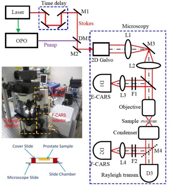
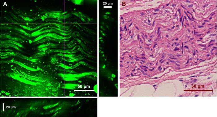
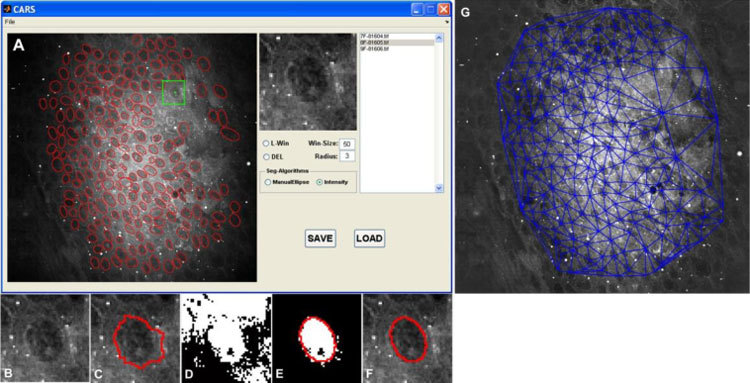
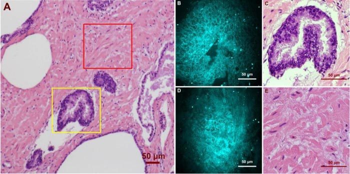
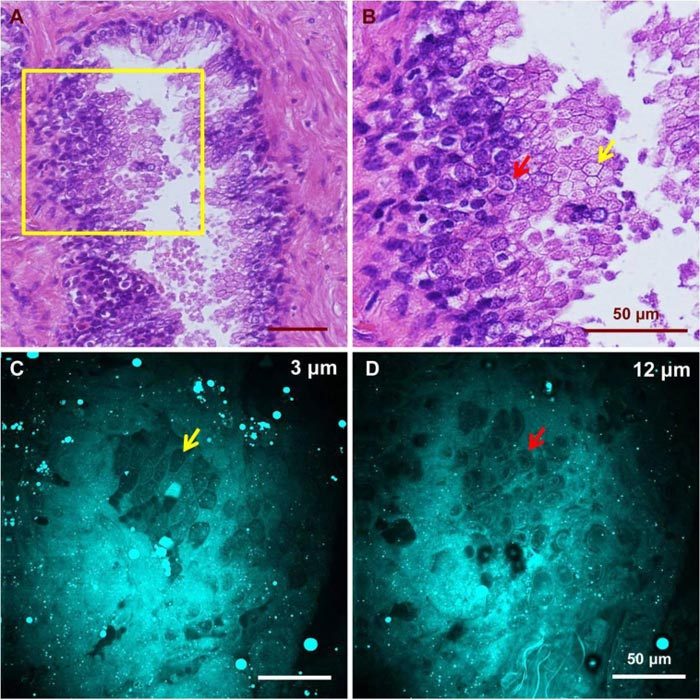
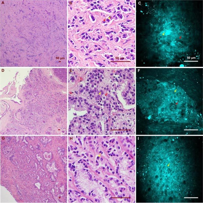
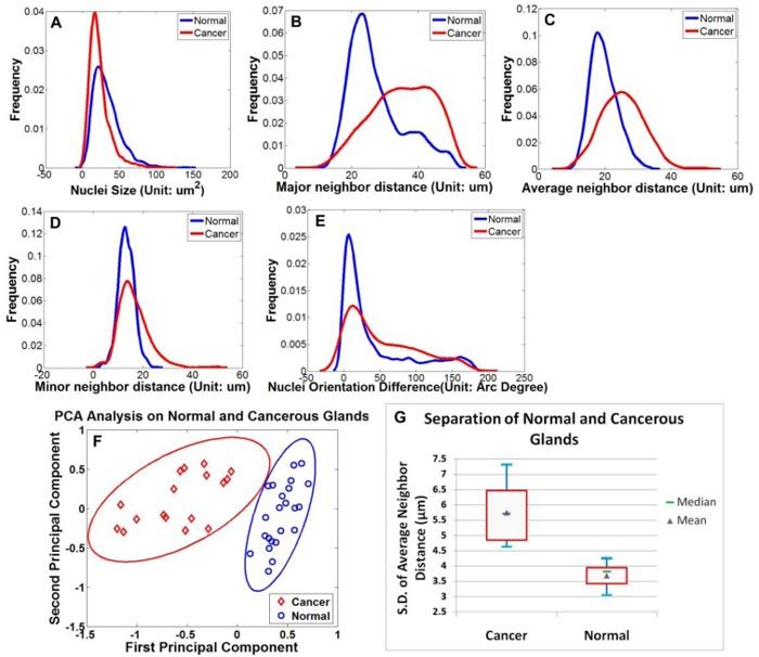
A custom built coherent anti-Stokes Raman scattering (CARS) microscope was used to image prostatic glands and nerve structures from 17 patients undergoing radical prostatectomy. Imaging of glandular and nerve structures showed distinctive cellular features that correlated to histological stains. Segmentation of cell nucleus was performed to establish a cell feature-based model to separate normal glands from cancer glands. In this study, we use a single parameter, average cell neighbor distance based on CARS imaging, to characterize normal and cancerous glandular structures. By combining CARS with our novel classification model, we are able to characterize prostate glandular and nerve structures in a manner that potentially enables real-time, intra-operative assessment of surgical margins and neurovascular bundles. As such, this method could potentially improve outcomes following radical prostatectomy.

摘要

使用一台定制的相干反斯托克斯拉曼散射(CARS)显微镜对17例接受根治性前列腺切除术患者的前列腺腺体和神经结构进行成像。腺体和神经结构成像显示出与组织学染色相关的独特细胞特征。进行细胞核分割以建立基于细胞特征的模型,将正常腺体与癌性腺体区分开来。在本研究中,我们使用基于CARS成像的单个参数——平均细胞邻近距离,来表征正常和癌性腺体结构。通过将CARS与我们的新型分类模型相结合,我们能够以一种潜在地实现手术切缘和神经血管束实时术中评估的方式来表征前列腺腺体和神经结构。因此,该方法可能会改善根治性前列腺切除术后的结果。

https://cdn.ncbi.nlm.nih.gov/pmc/blobs/5535/3072130/88762f02a4bd/boe-2-4-915-g007.jpg
https://cdn.ncbi.nlm.nih.gov/pmc/blobs/5535/3072130/8db7a479bc8b/boe-2-4-915-g001.jpg
https://cdn.ncbi.nlm.nih.gov/pmc/blobs/5535/3072130/66641b41ba5b/boe-2-4-915-g005.jpg
https://cdn.ncbi.nlm.nih.gov/pmc/blobs/5535/3072130/1e8a96a716fc/boe-2-4-915-g002.jpg
https://cdn.ncbi.nlm.nih.gov/pmc/blobs/5535/3072130/fe2c7e9f47f8/boe-2-4-915-g003.jpg
https://cdn.ncbi.nlm.nih.gov/pmc/blobs/5535/3072130/70c22a487494/boe-2-4-915-g004.jpg
https://cdn.ncbi.nlm.nih.gov/pmc/blobs/5535/3072130/06f273571061/boe-2-4-915-g006.jpg
https://cdn.ncbi.nlm.nih.gov/pmc/blobs/5535/3072130/88762f02a4bd/boe-2-4-915-g007.jpg

相似文献

1
Label-free high-resolution imaging of prostate glands and cavernous nerves using coherent anti-Stokes Raman scattering microscopy.使用相干反斯托克斯拉曼散射显微镜对前列腺和海绵体神经进行无标记高分辨率成像。
Biomed Opt Express. 2011 Mar 18;2(4):915-26. doi: 10.1364/BOE.2.000915.
2
Nerve Segmentation with Deep Learning from Label-Free Endoscopic Images Obtained Using Coherent Anti-Stokes Raman Scattering.基于相干反斯托克斯拉曼散射获得的无标记内窥镜图像的深度学习神经分割。
Biomolecules. 2020 Jul 8;10(7):1012. doi: 10.3390/biom10071012.
3
Implementation of a Coherent Anti-Stokes Raman Scattering (CARS) System on a Ti:Sapphire and OPO Laser Based Standard Laser Scanning Microscope.基于钛宝石和光学参量振荡器(OPO)激光器的标准激光扫描显微镜上的相干反斯托克斯拉曼散射(CARS)系统的实现
J Vis Exp. 2016 Jul 17(113). doi: 10.3791/54262.
4
Raman and coherent anti-Stokes Raman scattering microspectroscopy for biomedical applications.用于生物医学应用的喇曼和相干反斯托克斯拉曼散射微光谱学。
J Biomed Opt. 2012 Apr;17(4):040801. doi: 10.1117/1.JBO.17.4.040801.
5
Chemical fingerprinting of single glandular trichomes of Cannabis sativa by Coherent anti-Stokes Raman scattering (CARS) microscopy.利用相干反斯托克斯拉曼散射(CARS)显微镜对大麻单腺毛的化学指纹图谱进行分析。
BMC Plant Biol. 2018 Nov 12;18(1):275. doi: 10.1186/s12870-018-1481-4.
6
Vibrational spectroscopy and imaging with non-resonant coherent anti-Stokes Raman scattering: double stimulated Raman scattering scheme.非共振相干反斯托克斯拉曼散射的振动光谱学与成像:双受激拉曼散射方案
Opt Express. 2019 Aug 5;27(16):23558-23575. doi: 10.1364/OE.27.023558.
7
Quantitative chemical imaging with background-free multiplex coherent anti-Stokes Raman scattering by dual-soliton Stokes pulses.利用双孤子斯托克斯脉冲进行无背景多重相干反斯托克斯拉曼散射的定量化学成像。
Biomed Opt Express. 2016 Sep 9;7(10):3927-3939. doi: 10.1364/BOE.7.003927. eCollection 2016 Oct 1.
8
Texture analysis and classification in coherent anti-Stokes Raman scattering (CARS) microscopy images for automated detection of skin cancer.相干反斯托克斯拉曼散射(CARS)显微镜图像中的纹理分析和分类,用于皮肤癌的自动检测。
Comput Med Imaging Graph. 2015 Jul;43:36-43. doi: 10.1016/j.compmedimag.2015.02.010. Epub 2015 Mar 7.
9
Expanding multimodal microscopy by high spectral resolution coherent anti-Stokes Raman scattering imaging for clinical disease diagnostics.通过高光谱分辨率相干反斯托克斯拉曼散射成像扩展多模态显微镜,用于临床疾病诊断。
Anal Chem. 2013 Jul 16;85(14):6703-15. doi: 10.1021/ac400570w. Epub 2013 Jul 2.
10
Differential diagnosis of breast cancer using quantitative, label-free and molecular vibrational imaging.使用定量、无标记和分子振动成像技术对乳腺癌进行鉴别诊断。
Biomed Opt Express. 2011 Aug 1;2(8):2160-74. doi: 10.1364/BOE.2.002160. Epub 2011 Jul 5.

引用本文的文献

1
Vibrational spectroscopy and multiphoton microscopy for label-free visualization of nervous system degeneration and regeneration.振动光谱和多光子显微镜用于神经系统退变和再生的无标记可视化
Biophys Rev. 2023 Oct 5;16(2):219-235. doi: 10.1007/s12551-023-01158-2. eCollection 2024 Apr.
2
Raman difference spectroscopy and U-Net convolutional neural network for molecular analysis of cutaneous neurofibroma.拉曼差分光谱和 U-Net 卷积神经网络在皮肤神经纤维瘤的分子分析中的应用。
PLoS One. 2024 Apr 11;19(4):e0302017. doi: 10.1371/journal.pone.0302017. eCollection 2024.
3
Image-to-image translation of label-free molecular vibrational images for a histopathological review using the UNet+/seg-cGAN model.

本文引用的文献

1
Label-free cellular imaging by broadband coherent anti-Stokes Raman scattering microscopy.无标记细胞成像的宽带相干反斯托克斯拉曼散射显微镜。
Biophys J. 2010 Oct 20;99(8):2695-704. doi: 10.1016/j.bpj.2010.08.009.
2
Raman spectroscopic imaging for in vivo detection of cerebral brain metastases.拉曼光谱成像用于脑转移瘤的体内检测。
Anal Bioanal Chem. 2010 Oct;398(4):1707-13. doi: 10.1007/s00216-010-4116-7. Epub 2010 Aug 24.
3
Coherent anti-stokes Raman scattering microscopy: chemical imaging for biology and medicine.相干反斯托克斯拉曼散射显微镜:生物学和医学中的化学成像。
使用UNet+/seg-cGAN模型对无标记分子振动图像进行图像到图像的转换,用于组织病理学检查。
Biomed Opt Express. 2022 Mar 8;13(4):1924-1938. doi: 10.1364/BOE.445319. eCollection 2022 Apr 1.
4
Artificial intelligence-augmented, label-free molecular imaging method for tissue identification, cancer diagnosis, and cancer margin detection.用于组织识别、癌症诊断和癌症边缘检测的人工智能增强型无标记分子成像方法。
Biomed Opt Express. 2021 Aug 13;12(9):5559-5582. doi: 10.1364/BOE.428738. eCollection 2021 Sep 1.
5
Novel endoscopic optical diagnostic technologies in medical trial research: recent advancements and future prospects.新型内镜光学诊断技术在临床试验研究中的应用:最新进展与未来展望。
Biomed Eng Online. 2021 Jan 6;20(1):5. doi: 10.1186/s12938-020-00845-5.
6
Simultaneous label-free live imaging of cell nucleus and luminescent nanodiamonds.同时对细胞核和发光纳米金刚石进行无标记实时成像。
Sci Rep. 2020 Jun 17;10(1):9791. doi: 10.1038/s41598-020-66593-7.
7
Quantitative imaging of lipid droplets in single cells.单细胞中脂滴的定量成像。
Analyst. 2019 Jan 28;144(3):753-765. doi: 10.1039/c8an01525b.
8
Coherent anti-Stokes Raman scattering rigid endoscope toward robot-assisted surgery.用于机器人辅助手术的相干反斯托克斯拉曼散射刚性内窥镜。
Biomed Opt Express. 2018 Jan 2;9(2):387-396. doi: 10.1364/BOE.9.000387. eCollection 2018 Feb 1.
9
Combining deep learning and coherent anti-Stokes Raman scattering imaging for automated differential diagnosis of lung cancer.深度学习与相干反斯托克斯拉曼散射成像相结合,用于肺癌的自动鉴别诊断。
J Biomed Opt. 2017 Oct;22(10):1-10. doi: 10.1117/1.JBO.22.10.106017.
10
Rapid and accurate peripheral nerve imaging by multipoint Raman spectroscopy.多点拉曼光谱法快速准确的外周神经成像。
Sci Rep. 2017 Apr 12;7(1):845. doi: 10.1038/s41598-017-00995-y.
Annu Rev Anal Chem (Palo Alto Calif). 2008;1:883-909. doi: 10.1146/annurev.anchem.1.031207.112754.
4
Delivery of picosecond lasers in multimode fibers for coherent anti-Stokes Raman scattering imaging.用于相干反斯托克斯拉曼散射成像的多模光纤中皮秒激光的传输。
Opt Express. 2010 Jun 7;18(12):13017-28. doi: 10.1364/OE.18.013017.
5
Fiber delivered probe for efficient CARS imaging of tissues.用于组织高效相干反斯托克斯拉曼散射(CARS)成像的光纤传输探头。
Opt Express. 2010 Feb 1;18(3):2380-8. doi: 10.1364/OE.18.002380.
6
Variability in diagnostic opinion among pathologists for single small atypical foci in prostate biopsies.前列腺穿刺活检中单发小灶不典型时病理医师诊断意见的差异。
Am J Surg Pathol. 2010 Feb;34(2):169-77. doi: 10.1097/PAS.0b013e3181c7997b.
7
Robot-assisted radical prostatectomy: advances since 2005.机器人辅助根治性前列腺切除术:2005 年以来的进展。
Curr Opin Urol. 2010 Mar;20(2):130-5. doi: 10.1097/MOU.0b013e328336257a.
8
Multiple nuclei tracking using integer programming for quantitative cancer cell cycle analysis.使用整数规划进行多核跟踪以进行定量癌细胞周期分析。
IEEE Trans Med Imaging. 2010 Jan;29(1):96-105. doi: 10.1109/TMI.2009.2027813. Epub 2009 Jul 28.
9
Towards CARS Endoscopy.迈向相干反斯托克斯拉曼散射(CARS)内窥镜检查
Opt Express. 2006 May 15;14(10):4427-32. doi: 10.1364/oe.14.004427.
10
Multiphoton microscopy of prostate and periprostatic neural tissue: a promising imaging technique for improving nerve-sparing prostatectomy.前列腺及前列腺周围神经组织的多光子显微镜检查:一种用于改进保留神经前列腺切除术的有前景的成像技术。
J Endourol. 2009 May;23(5):861-7. doi: 10.1089/end.2009.0221.