Translational Biophotonics Laboratory, Department of Systems Medicine and Bioengineering, Houston Me, United States.
Rice University, Department of Electrical and Computer Engineering, Houston, Texas, United States.
J Biomed Opt. 2017 Oct;22(10):1-10. doi: 10.1117/1.JBO.22.10.106017.
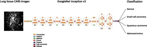
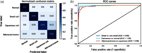
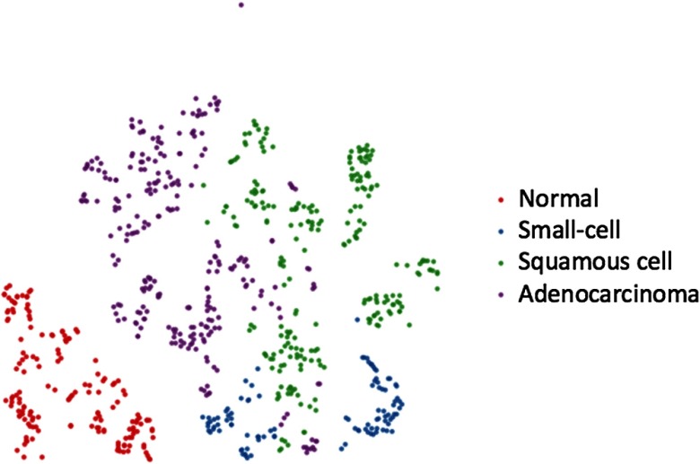
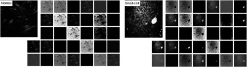
Lung cancer is the most prevalent type of cancer and the leading cause of cancer-related deaths worldwide. Coherent anti-Stokes Raman scattering (CARS) is capable of providing cellular-level images and resolving pathologically related features on human lung tissues. However, conventional means of analyzing CARS images requires extensive image processing, feature engineering, and human intervention. This study demonstrates the feasibility of applying a deep learning algorithm to automatically differentiate normal and cancerous lung tissue images acquired by CARS. We leverage the features learned by pretrained deep neural networks and retrain the model using CARS images as the input. We achieve 89.2% accuracy in classifying normal, small-cell carcinoma, adenocarcinoma, and squamous cell carcinoma lung images. This computational method is a step toward on-the-spot diagnosis of lung cancer and can be further strengthened by the efforts aimed at miniaturizing the CARS technique for fiber-based microendoscopic imaging.
肺癌是最常见的癌症类型,也是全球癌症相关死亡的主要原因。相干反斯托克斯拉曼散射(CARS)能够提供细胞水平的图像,并解析人类肺部组织的病理相关特征。然而,传统的 CARS 图像分析方法需要大量的图像处理、特征工程和人工干预。本研究展示了应用深度学习算法自动区分 CARS 采集的正常和癌变肺组织图像的可行性。我们利用预训练的深度神经网络所学习到的特征,并使用 CARS 图像作为输入重新训练模型。我们在分类正常、小细胞癌、腺癌和鳞状细胞癌肺图像方面取得了 89.2%的准确率。这种计算方法是实现肺癌现场诊断的重要一步,并可以通过努力进一步加强,即将 CARS 技术小型化用于基于光纤的微内窥镜成像。