Department of Pathology and Laboratory Medicine, Alpert Medical School of Brown University, Providence, Rhode Island, United States of America.
PLoS One. 2011 Mar 31;6(3):e17966. doi: 10.1371/journal.pone.0017966.
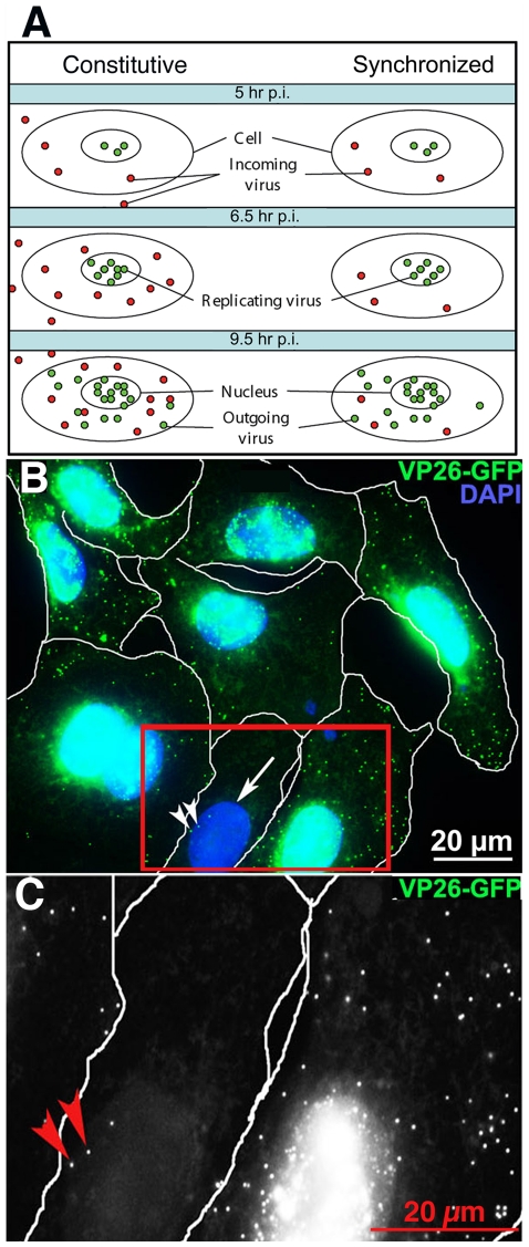
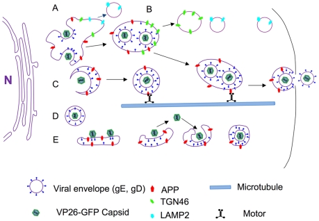
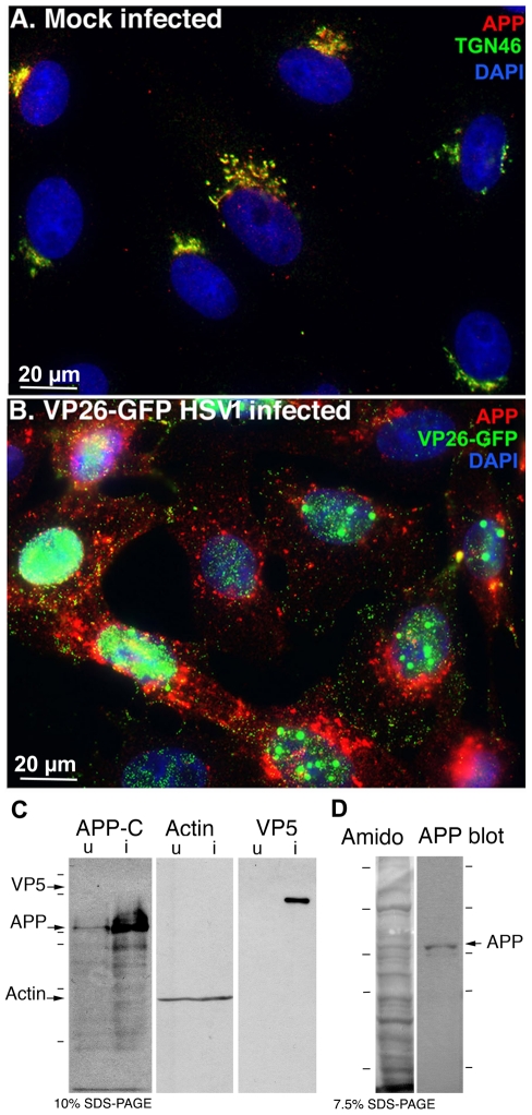
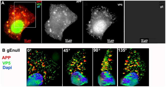
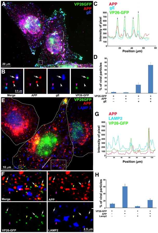
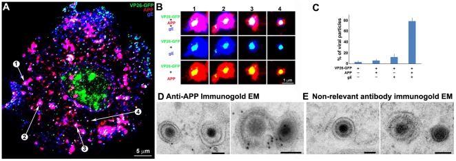
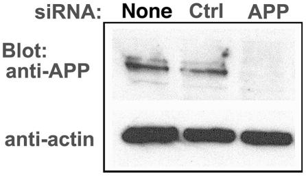
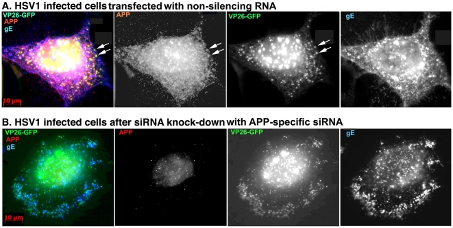
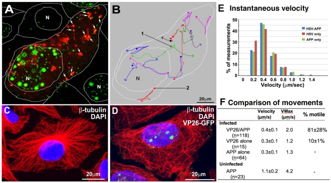
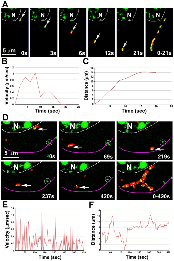
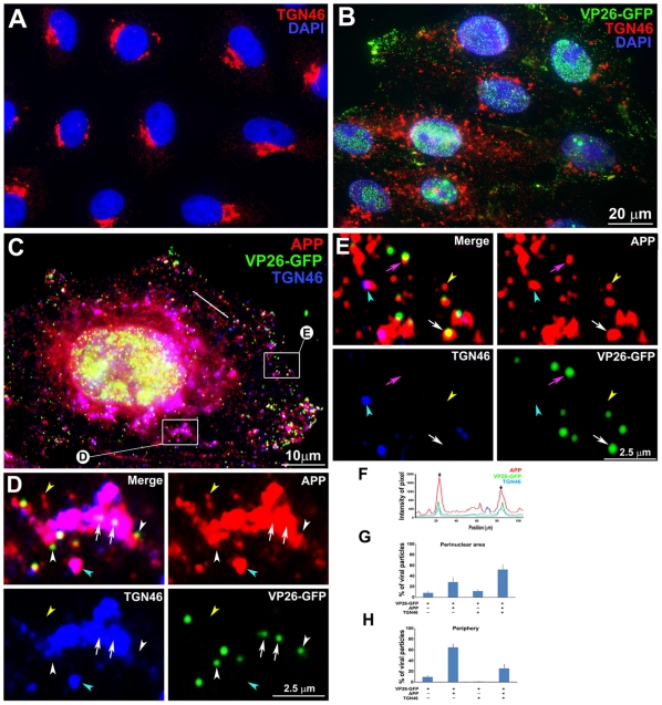
Herpes simplex type 1 (HSV1) replicates in epithelial cells and secondarily enters local sensory neuronal processes, traveling retrograde to the neuronal nucleus to enter latency. Upon reawakening newly synthesized viral particles travel anterograde back to the epithelial cells of the lip, causing the recurrent cold sore. HSV1 co-purifies with amyloid precursor protein (APP), a cellular transmembrane glycoprotein and receptor for anterograde transport machinery that when proteolyzed produces A-beta, the major component of senile plaques. Here we focus on transport inside epithelial cells of newly synthesized virus during its transit to the cell surface. We hypothesize that HSV1 recruits cellular APP during transport. We explore this with quantitative immuno-fluorescence, immuno-gold electron-microscopy and live cell confocal imaging. After synchronous infection most nascent VP26-GFP-labeled viral particles in the cytoplasm co-localize with APP (72.8+/-6.7%) and travel together with APP inside living cells (81.1+/-28.9%). This interaction has functional consequences: HSV1 infection decreases the average velocity of APP particles (from 1.1+/-0.2 to 0.3+/-0.1 µm/s) and results in APP mal-distribution in infected cells, while interplay with APP-particles increases the frequency (from 10% to 81% motile) and velocity (from 0.3+/-0.1 to 0.4+/-0.1 µm/s) of VP26-GFP transport. In cells infected with HSV1 lacking the viral Fc receptor, gE, an envelope glycoprotein also involved in viral axonal transport, APP-capsid interactions are preserved while the distribution and dynamics of dual-label particles differ from wild-type by both immuno-fluorescence and live imaging. Knock-down of APP with siRNA eliminates APP staining, confirming specificity. Our results indicate that most intracellular HSV1 particles undergo frequent dynamic interplay with APP in a manner that facilitates viral transport and interferes with normal APP transport and distribution. Such dynamic interactions between APP and HSV1 suggest a mechanistic basis for the observed clinical relationship between HSV1 seropositivity and risk of Alzheimer's disease.
单纯疱疹病毒 1 型(HSV1)在上皮细胞中复制,然后进入局部感觉神经元的过程,逆行进入神经元核进入潜伏期。当重新唤醒时,新合成的病毒颗粒向前逆行回到唇部的上皮细胞,引起复发性唇疱疹。HSV1 与淀粉样前体蛋白(APP)共纯化,APP 是一种细胞跨膜糖蛋白,也是顺行转运机制的受体,当被蛋白水解时会产生 A-β,这是老年斑的主要成分。在这里,我们专注于新合成病毒在转移到细胞表面过程中穿过上皮细胞的内部运输。我们假设 HSV1 在运输过程中招募细胞 APP。我们通过定量免疫荧光、免疫金电子显微镜和活细胞共聚焦成像来探索这一点。在同步感染后,细胞质中大多数新出现的 VP26-GFP 标记的病毒颗粒与 APP 共定位(72.8+/-6.7%),并与 APP 一起在活细胞内运输(81.1+/-28.9%)。这种相互作用具有功能后果:HSV1 感染降低了 APP 颗粒的平均速度(从 1.1+/-0.2 到 0.3+/-0.1 µm/s),并导致感染细胞中 APP 的分布异常,而与 APP 颗粒的相互作用增加了 VP26-GFP 运输的频率(从 10%到 81%运动)和速度(从 0.3+/-0.1 到 0.4+/-0.1 µm/s)。在感染缺乏病毒 Fc 受体 gE 的 HSV1 的细胞中,包膜糖蛋白也参与病毒轴突运输,与衣壳的 APP 相互作用得以保留,而双标记颗粒的分布和动力学通过免疫荧光和活细胞成像与野生型不同。用 siRNA 敲低 APP 消除了 APP 染色,证实了特异性。我们的结果表明,大多数细胞内 HSV1 颗粒与 APP 频繁地进行动态相互作用,这促进了病毒的运输,并干扰了正常的 APP 运输和分布。APP 与 HSV1 之间的这种动态相互作用表明了观察到的 HSV1 血清阳性与阿尔茨海默病风险之间的临床关系的机制基础。