Department of Clinical Physiology, Lund University, Skåne University Hospital Lund, Lund, Sweden.
BMC Med Imaging. 2011 Apr 12;11:10. doi: 10.1186/1471-2342-11-10.
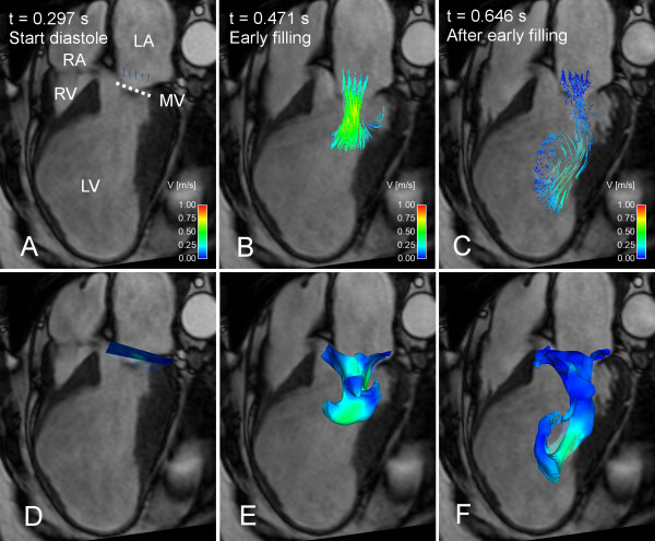
Functional and morphological changes of the heart influence blood flow patterns. Therefore, flow patterns may carry diagnostic and prognostic information. Three-dimensional, time-resolved, three-directional phase contrast cardiovascular magnetic resonance (4D PC-CMR) can image flow patterns with unique detail, and using new flow visualization methods may lead to new insights. The aim of this study is to present and validate a novel visualization method with a quantitative potential for blood flow from 4D PC-CMR, called Volume Tracking, and investigate if Volume Tracking complements particle tracing, the most common visualization method used today.
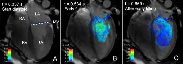
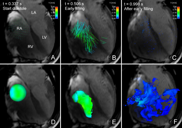
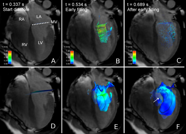
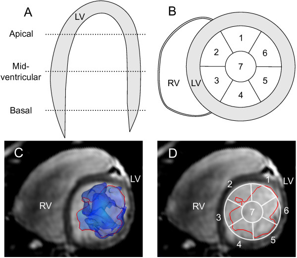
Eight healthy volunteers and one patient with a large apical left ventricular aneurysm underwent 4D PC-CMR flow imaging of the whole heart. Volume Tracking and particle tracing visualizations were compared visually side-by-side in a visualization software package. To validate Volume Tracking, the number of particle traces that agreed with the Volume Tracking visualizations was counted and expressed as a percentage of total released particles in mid-diastole and end-diastole respectively. Two independent observers described blood flow patterns in the left ventricle using Volume Tracking visualizations.
Volume Tracking was feasible in all eight healthy volunteers and in the patient. Visually, Volume Tracking and particle tracing are complementary methods, showing different aspects of the flow. When validated against particle tracing, on average 90.5% and 87.8% of the particles agreed with the Volume Tracking surface in mid-diastole and end-diastole respectively. Inflow patterns in the left ventricle varied between the subjects, with excellent agreement between observers. The left ventricular inflow pattern in the patient differed from the healthy subjects.
Volume Tracking is a new visualization method for blood flow measured by 4D PC-CMR. Volume Tracking complements and provides incremental information compared to particle tracing that may lead to a better understanding of blood flow and may improve diagnosis and prognosis of cardiovascular diseases.
心脏的功能和形态变化会影响血流模式。因此,血流模式可能具有诊断和预后信息。三维、时间分辨、三方向相位对比心血管磁共振(4D PC-CMR)可以用独特的细节成像血流模式,使用新的血流可视化方法可能会带来新的见解。本研究的目的是介绍和验证一种新的可视化方法,该方法具有 4D PC-CMR 血流的定量潜力,称为容积追踪,并研究容积追踪是否补充了当今最常用的可视化方法——粒子追踪。
8 名健康志愿者和 1 名患有巨大心尖左心室瘤的患者接受了整个心脏的 4D PC-CMR 血流成像。在可视化软件包中,并排比较容积追踪和粒子追踪的可视化效果。为了验证容积追踪,计数与容积追踪可视化结果一致的粒子轨迹数量,并分别表示为中舒张期和舒张末期释放的总粒子数的百分比。两名独立观察者使用容积追踪可视化描述左心室的血流模式。
容积追踪在所有 8 名健康志愿者和患者中均可行。从视觉上看,容积追踪和粒子追踪是互补的方法,显示了血流的不同方面。与粒子追踪进行验证时,平均有 90.5%和 87.8%的粒子分别在中舒张期和舒张末期与容积追踪表面一致。左心室流入模式在受试者之间存在差异,观察者之间具有极好的一致性。患者的左心室流入模式与健康受试者不同。
容积追踪是一种新的可视化方法,用于测量 4D PC-CMR 的血流。与粒子追踪相比,容积追踪具有互补性并提供增量信息,这可能会更好地理解血流,并可能改善心血管疾病的诊断和预后。