• 文献检索
  • 文档翻译
  • 深度研究
  • 学术资讯
  • Suppr Zotero 插件Zotero 插件
  • 邀请有礼
  • 套餐&价格
  • 历史记录
应用&插件
Suppr Zotero 插件Zotero 插件浏览器插件Mac 客户端Windows 客户端微信小程序
定价
高级版会员购买积分包购买API积分包
服务
文献检索文档翻译深度研究API 文档MCP 服务
关于我们
关于 Suppr公司介绍联系我们用户协议隐私条款
关注我们

Suppr 超能文献

核心技术专利:CN118964589B侵权必究
粤ICP备2023148730 号-1Suppr @ 2026

文献检索

告别复杂PubMed语法,用中文像聊天一样搜索,搜遍4000万医学文献。AI智能推荐,让科研检索更轻松。

立即免费搜索

文件翻译

保留排版,准确专业,支持PDF/Word/PPT等文件格式,支持 12+语言互译。

免费翻译文档

深度研究

AI帮你快速写综述,25分钟生成高质量综述,智能提取关键信息,辅助科研写作。

立即免费体验

健康受试者左心室 4D 血流心血管磁共振测量的重测变异性。

Test-retest variability of left ventricular 4D flow cardiovascular magnetic resonance measurements in healthy subjects.

机构信息

University of Oxford Centre for Clinical Magnetic Resonance Research (OCMR), Division of Cardiovascular Medicine, Radcliffe Department of Medicine, Oxford, UK.

Division of Cardiovascular Medicine, Linköping University, Linköping, Sweden.

出版信息

J Cardiovasc Magn Reson. 2018 Mar 2;20(1):15. doi: 10.1186/s12968-018-0432-4.

DOI:10.1186/s12968-018-0432-4
PMID:29499706
原文链接:https://pmc.ncbi.nlm.nih.gov/articles/PMC5833126/
Abstract

BACKGROUND

Quantification and visualisation of left ventricular (LV) blood flow is afforded by three-dimensional, time resolved phase contrast cardiovascular magnetic resonance (CMR 4D flow). However, few data exist upon the repeatability and variability of these parameters in a healthy population. We aimed to assess the repeatability and variability over time of LV 4D CMR flow measurements.

METHODS

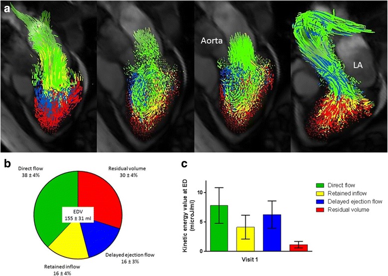
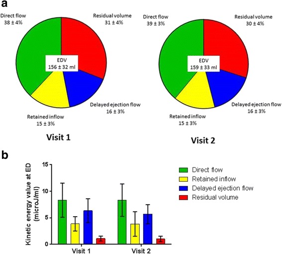
Forty five controls underwent CMR 4D flow data acquisition. Of these, 10 underwent a second scan within the same visit (scan-rescan), 25 returned for a second visit (interval scan; median interval 52 days, IQR 28-57 days). The LV-end diastolic volume (EDV) was divided into four flow components: 1) Direct flow: inflow that passes directly to ejection; 2) Retained inflow: inflow that enters and resides within the LV; 3) Delayed ejection flow: starts within the LV and is ejected and 4) Residual volume: blood that resides within the LV for > 2 cardiac cycles. Each flow components' volume was related to the EDV (volume-ratio). The kinetic energy at end-diastole (ED) was measured and divided by the components' volume.

RESULTS

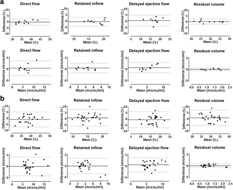
The dominant flow component in all 45 controls was the direct flow (volume ratio 38 ± 4%) followed by the residual volume (30 ± 4%), then delayed ejection flow (16 ± 3%) and retained inflow (16 ± 4%). The kinetic energy at ED for each component was direct flow (7.8 ± 3.0 microJ/ml), retained inflow (4.1 ± 2.0 microJ/ml), delayed ejection flow (6.3 ± 2.3 microJ/ml) and the residual volume (1.2 ± 0.5 microJ/ml). The coefficients of variation for the scan-rescan ranged from 2.5%-9.2% for the flow components' volume ratio and between 13.5%-17.7% for the kinetic energy. The interval scan results showed higher coefficients of variation with values from 6.2-16.1% for the flow components' volume ratio and 16.9-29.0% for the kinetic energy of the flow components.

CONCLUSION

LV flow components' volume and their associated kinetic energy values are repeatable and stable within a population over time. However, the variability of these measurements in individuals over time is greater than can be attributed to sources of error in the data acquisition and analysis, suggesting that additional physiological factors may influence LV flow measurements.

摘要

背景

三维时间分辨相位对比心血管磁共振(CMR 4D 流)可定量和可视化左心室(LV)血流。然而,在健康人群中,关于这些参数的重复性和可变性的数据很少。我们旨在评估 LV 4D CMR 流测量的重复性和随时间的可变性。

方法

45 名对照者接受 CMR 4D 流数据采集。其中 10 名在同一就诊期间进行第二次扫描(扫描-重扫),25 名在第二次就诊时返回(间隔扫描;中位间隔 52 天,IQR 28-57 天)。LV 舒张末期容积(EDV)分为四个血流成分:1)直接血流:直接流入射血;2)滞留的流入:流入并驻留在 LV 内;3)延迟射流:开始于 LV 内并被射出;4)残余容积:在 LV 内停留超过 2 个心动周期的血液。每个血流成分的体积与 EDV 相关(体积比)。在舒张末期(ED)测量动能,并除以各成分的体积。

结果

所有 45 名对照者的主要血流成分均为直接血流(体积比 38±4%),其次为残余容积(30±4%),然后为延迟射流(16±3%)和滞留的流入(16±4%)。每个成分在 ED 的动能分别为直接血流(7.8±3.0 微焦耳/毫升)、滞留的流入(4.1±2.0 微焦耳/毫升)、延迟射流(6.3±2.3 微焦耳/毫升)和残余容积(1.2±0.5 微焦耳/毫升)。扫描-重扫的变异系数为血流成分体积比的 2.5%-9.2%,动能的 13.5%-17.7%。间隔扫描结果显示,血流成分体积比的变异系数较高,为 6.2-16.1%,动能的变异系数为 16.9-29.0%。

结论

LV 血流成分的体积及其相关动能值在人群中随时间具有重复性和稳定性。然而,个体随时间的这些测量值的可变性大于可以归因于数据采集和分析中的误差源,这表明其他生理因素可能会影响 LV 血流测量。

https://cdn.ncbi.nlm.nih.gov/pmc/blobs/3a53/5833126/c9cca3193bb6/12968_2018_432_Fig3_HTML.jpg
https://cdn.ncbi.nlm.nih.gov/pmc/blobs/3a53/5833126/4f5a51a80a2f/12968_2018_432_Fig1_HTML.jpg
https://cdn.ncbi.nlm.nih.gov/pmc/blobs/3a53/5833126/5e98ed1bef62/12968_2018_432_Fig2_HTML.jpg
https://cdn.ncbi.nlm.nih.gov/pmc/blobs/3a53/5833126/c9cca3193bb6/12968_2018_432_Fig3_HTML.jpg
https://cdn.ncbi.nlm.nih.gov/pmc/blobs/3a53/5833126/4f5a51a80a2f/12968_2018_432_Fig1_HTML.jpg
https://cdn.ncbi.nlm.nih.gov/pmc/blobs/3a53/5833126/5e98ed1bef62/12968_2018_432_Fig2_HTML.jpg
https://cdn.ncbi.nlm.nih.gov/pmc/blobs/3a53/5833126/c9cca3193bb6/12968_2018_432_Fig3_HTML.jpg

相似文献

1
Test-retest variability of left ventricular 4D flow cardiovascular magnetic resonance measurements in healthy subjects.健康受试者左心室 4D 血流心血管磁共振测量的重测变异性。
J Cardiovasc Magn Reson. 2018 Mar 2;20(1):15. doi: 10.1186/s12968-018-0432-4.
2
Impact of age, sex and ethnicity on intra-cardiac flow components and left ventricular kinetic energy derived from 4D flow CMR.年龄、性别和种族对源自4D流心脏磁共振成像的心脏内血流成分和左心室动能的影响。
Int J Cardiol. 2021 Aug 1;336:105-112. doi: 10.1016/j.ijcard.2021.05.035. Epub 2021 May 25.
3
Mechanical dyssynchrony alters left ventricular flow energetics in failing hearts with LBBB: a 4D flow CMR pilot study.机械性不同步改变左束支传导阻滞所致心力衰竭患者左心室血流能量学:一项四维血流心脏磁共振成像初步研究。
Int J Cardiovasc Imaging. 2018 Apr;34(4):587-596. doi: 10.1007/s10554-017-1261-5. Epub 2017 Nov 2.
4
Deep learning based automated left ventricle segmentation and flow quantification in 4D flow cardiac MRI.基于深度学习的 4D 流心脏 MRI 中左心室自动分割和流量定量
J Cardiovasc Magn Reson. 2024 Summer;26(1):100003. doi: 10.1016/j.jocmr.2023.100003. Epub 2024 Jan 10.
5
Semi-automatic quantification of 4D left ventricular blood flow.四维左心室血流半自动定量。
J Cardiovasc Magn Reson. 2010 Feb 12;12(1):9. doi: 10.1186/1532-429X-12-9.
6
Scan-rescan reproducibility of diastolic left ventricular kinetic energy, viscous energy loss and vorticity assessment using 4D flow MRI: analysis in healthy subjects.使用4D流磁共振成像评估舒张期左心室动能、粘性能量损失和涡度的扫描-再扫描重复性:健康受试者分析
Int J Cardiovasc Imaging. 2018 Jun;34(6):905-920. doi: 10.1007/s10554-017-1291-z. Epub 2018 Jan 5.
7
Left ventricular blood flow kinetic energy after myocardial infarction - insights from 4D flow cardiovascular magnetic resonance.心肌梗死后左心室血流动能 - 4D 流心血管磁共振的见解。
J Cardiovasc Magn Reson. 2018 Aug 30;20(1):61. doi: 10.1186/s12968-018-0483-6.
8
Age- and sex-specific reference values of biventricular flow components and kinetic energy by 4D flow cardiovascular magnetic resonance in healthy subjects.健康受试者四维血流心血管磁共振左、右心室血流成分和动能的年龄和性别特异性参考值。
J Cardiovasc Magn Reson. 2023 Sep 18;25(1):50. doi: 10.1186/s12968-023-00960-x.
9
Ventricular flow analysis and its association with exertional capacity in repaired tetralogy of Fallot: 4D flow cardiovascular magnetic resonance study.修复后法洛四联症的心室血流分析及其与运动能力的相关性:4D 流心血管磁共振研究。
J Cardiovasc Magn Reson. 2022 Jan 3;24(1):4. doi: 10.1186/s12968-021-00832-2.
10
Evaluation of Left Ventricular Flow Kinetic Energy by Four-Dimensional Blood Flow MRI in Nondialysis Chronic Kidney Disease Patients.非透析慢性肾脏病患者左心室血流动能的四维血流磁共振成像评估
J Magn Reson Imaging. 2025 Feb;61(2):865-879. doi: 10.1002/jmri.29435. Epub 2024 May 6.

引用本文的文献

1
Cardiovascular magnetic resonance left ventricular 4D-flow: differences in flow components and kinetic energy across heart failure spectrum.心血管磁共振左心室四维血流:心力衰竭谱中血流成分和动能的差异。
Eur Heart J Imaging Methods Pract. 2025 May 14;3(1):qyaf059. doi: 10.1093/ehjimp/qyaf059. eCollection 2025 Jan.
2
Diastolic Right Ventricle Dysfunction in Repaired Tetralogy of Fallot: Is it Time for 4D-Flow-MRI Parameters?法洛四联症修复术后的舒张期右心室功能障碍:是时候关注四维血流磁共振成像参数了吗?
JACC Asia. 2024 Dec 3;4(12):925-927. doi: 10.1016/j.jacasi.2024.10.018. eCollection 2024 Dec.
3
Observer- and sequence variability in personalized 4D flow MRI-based cardiovascular models.

本文引用的文献

1
Reference ranges for cardiac structure and function using cardiovascular magnetic resonance (CMR) in Caucasians from the UK Biobank population cohort.英国生物银行人群队列中白种人使用心血管磁共振成像(CMR)测量心脏结构和功能的参考范围
J Cardiovasc Magn Reson. 2017 Feb 3;19(1):18. doi: 10.1186/s12968-017-0327-9.
2
Altered Diastolic Flow Patterns and Kinetic Energy in Subtle Left Ventricular Remodeling and Dysfunction Detected by 4D Flow MRI.通过4D流MRI检测到的轻微左心室重构和功能障碍中的舒张期血流模式和动能改变
PLoS One. 2016 Aug 17;11(8):e0161391. doi: 10.1371/journal.pone.0161391. eCollection 2016.
3
Disturbed Intracardiac Flow Organization After Atrioventricular Septal Defect Correction as Assessed With 4D Flow Magnetic Resonance Imaging and Quantitative Particle Tracing.
基于个性化4D流磁共振成像的心血管模型中的观察者和序列变异性。
Sci Rep. 2025 Jan 8;15(1):1352. doi: 10.1038/s41598-024-84390-4.
4
Direct comparison of whole heart quantifications between different retrospective and prospective gated 4D flow CMR acquisitions.不同回顾性和前瞻性门控4D流CMR采集之间全心定量的直接比较。
Front Cardiovasc Med. 2024 Jul 31;11:1411752. doi: 10.3389/fcvm.2024.1411752. eCollection 2024.
5
Intraventricular 4D flow cardiovascular magnetic resonance for assessing patients with heart failure with preserved ejection fraction: a pilot study.用于评估射血分数保留的心力衰竭患者的脑室内四维血流心血管磁共振成像:一项初步研究。
Int J Cardiovasc Imaging. 2023 Oct;39(10):2015-2027. doi: 10.1007/s10554-023-02909-8. Epub 2023 Jun 28.
6
Intra-Cardiac Flow from Geometry Prescribed Computational Fluid Dynamics: Comparison with Ultrasound Vector Flow Imaging.心内血流的计算流体动力学:与超声向量流成像的比较。
Cardiovasc Eng Technol. 2023 Aug;14(4):489-504. doi: 10.1007/s13239-023-00666-2. Epub 2023 Jun 15.
7
Novel insights into diminished cardiac reserve in non-obstructive hypertrophic cardiomyopathy from four-dimensional flow cardiac magnetic resonance component analysis.从四维血流心脏磁共振分量分析看非梗阻性肥厚型心肌病心脏储备功能降低的新认识。
Eur Heart J Cardiovasc Imaging. 2023 Aug 23;24(9):1192-1200. doi: 10.1093/ehjci/jead074.
8
Impact of dobutamine stress on diastolic energetic efficiency of healthy left ventricle: an kinetic energy analysis.多巴酚丁胺负荷对健康左心室舒张能量效率的影响:一项动能分析
Front Cardiovasc Med. 2023 Mar 21;10:1103751. doi: 10.3389/fcvm.2023.1103751. eCollection 2023.
9
Visualizing diastolic failure: Non-invasive imaging-biomarkers in patients with heart failure with preserved ejection fraction.可视化舒张性心力衰竭:射血分数保留的心力衰竭患者的无创影像学生物标志物。
EBioMedicine. 2022 Dec;86:104369. doi: 10.1016/j.ebiom.2022.104369. Epub 2022 Nov 21.
10
Evaluation of Left Ventricular Function Using Four-Dimensional Flow Cardiovascular Magnetic Resonance: A Systematic Review.使用四维血流心血管磁共振评估左心室功能:一项系统评价。
J Cardiovasc Dev Dis. 2022 Sep 12;9(9):304. doi: 10.3390/jcdd9090304.
经4D流磁共振成像和定量粒子追踪评估,房室间隔缺损修复术后心内血流组织紊乱
Invest Radiol. 2015 Dec;50(12):850-7. doi: 10.1097/RLI.0000000000000194.
4
Variable velocity encoding in a three-dimensional, three-directional phase contrast sequence: Evaluation in phantom and volunteers.三维三方向相位对比序列中的可变速度编码:在体模和志愿者中的评估。
J Magn Reson Imaging. 2012 Dec;36(6):1450-9. doi: 10.1002/jmri.23778. Epub 2012 Oct 12.
5
Gender-specific differences in left ventricular remodelling in obesity: insights from cardiovascular magnetic resonance imaging.肥胖患者左心室重构的性别差异:心血管磁共振成像的见解。
Eur Heart J. 2013 Jan;34(4):292-9. doi: 10.1093/eurheartj/ehs341. Epub 2012 Oct 10.
6
Four-dimensional blood flow-specific markers of LV dysfunction in dilated cardiomyopathy.四维血流特异性标志物在扩张型心肌病中的左心室功能障碍。
Eur Heart J Cardiovasc Imaging. 2013 May;14(5):417-24. doi: 10.1093/ehjci/jes159. Epub 2012 Aug 8.
7
Quantification of presystolic blood flow organization and energetics in the human left ventricle.定量分析人体左心室收缩前期血流的组织和能量。
Am J Physiol Heart Circ Physiol. 2011 Jun;300(6):H2135-41. doi: 10.1152/ajpheart.00993.2010. Epub 2011 Mar 18.
8
Passing strange: flow in the failing ventricle.奇特现象:衰竭心室中的血流
Circ Heart Fail. 2010 Mar;3(2):326-31. doi: 10.1161/CIRCHEARTFAILURE.109.911867.
9
Semi-automatic quantification of 4D left ventricular blood flow.四维左心室血流半自动定量。
J Cardiovasc Magn Reson. 2010 Feb 12;12(1):9. doi: 10.1186/1532-429X-12-9.
10
Design and validation of Segment--freely available software for cardiovascular image analysis.设计和验证——用于心血管图像分析的免费软件段。
BMC Med Imaging. 2010 Jan 11;10:1. doi: 10.1186/1471-2342-10-1.