• 文献检索
  • 文档翻译
  • 深度研究
  • 学术资讯
  • Suppr Zotero 插件Zotero 插件
  • 邀请有礼
  • 套餐&价格
  • 历史记录
应用&插件
Suppr Zotero 插件Zotero 插件浏览器插件Mac 客户端Windows 客户端微信小程序
定价
高级版会员购买积分包购买API积分包
服务
文献检索文档翻译深度研究API 文档MCP 服务
关于我们
关于 Suppr公司介绍联系我们用户协议隐私条款
关注我们

Suppr 超能文献

核心技术专利:CN118964589B侵权必究
粤ICP备2023148730 号-1Suppr @ 2026

文献检索

告别复杂PubMed语法,用中文像聊天一样搜索,搜遍4000万医学文献。AI智能推荐,让科研检索更轻松。

立即免费搜索

文件翻译

保留排版,准确专业,支持PDF/Word/PPT等文件格式,支持 12+语言互译。

免费翻译文档

深度研究

AI帮你快速写综述,25分钟生成高质量综述,智能提取关键信息,辅助科研写作。

立即免费体验

四维左心室血流半自动定量。

Semi-automatic quantification of 4D left ventricular blood flow.

机构信息

Division of Cardiovascular Medicine, Department of Medical and Health Sciences, Linköping University, Linköping, Sweden.

出版信息

J Cardiovasc Magn Reson. 2010 Feb 12;12(1):9. doi: 10.1186/1532-429X-12-9.

DOI:10.1186/1532-429X-12-9
PMID:20152026
原文链接:https://pmc.ncbi.nlm.nih.gov/articles/PMC2831022/
Abstract

BACKGROUND

The beating heart is the generator of blood flow through the cardiovascular system. Within the heart's own chambers, normal complex blood flow patterns can be disturbed by diseases. Methods for the quantification of intra-cardiac blood flow, with its 4D (3D+time) nature, are lacking. We sought to develop and validate a novel semi-automatic analysis approach that integrates flow and morphological data.

METHOD

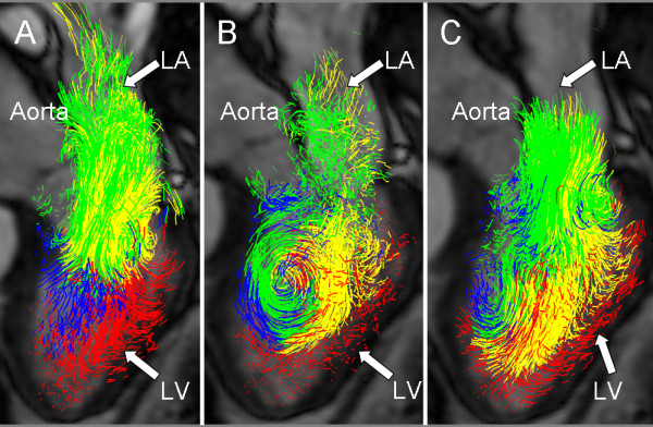
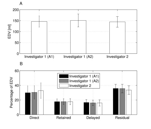
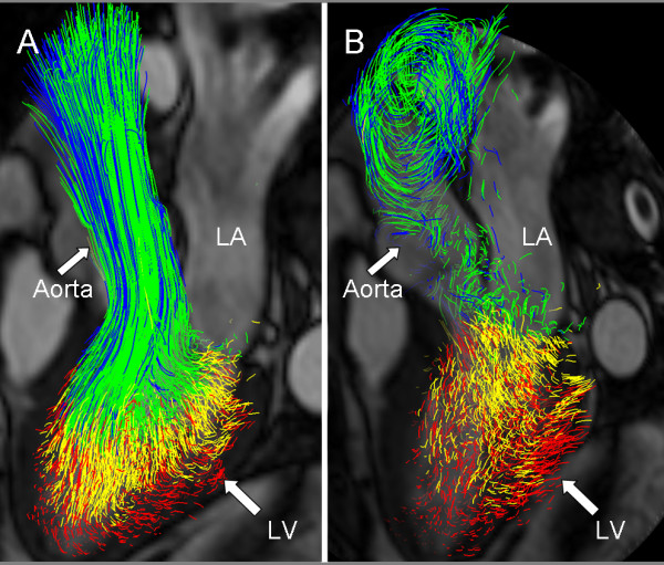
In six healthy subjects and three patients with dilated cardiomyopathy, three-directional, three-dimensional cine phase-contrast cardiovascular magnetic resonance (CMR) velocity data and balanced steady-state free-precession long- and short-axis images were acquired. The LV endocardium was segmented from the short-axis images at the times of isovolumetric contraction (IVC) and isovolumetric relaxation (IVR). At the time of IVC, pathlines were emitted from the IVC LV blood volume and traced forwards and backwards in time until IVR, thus including the entire cardiac cycle. The IVR volume was used to determine if and where the pathlines left the LV. This information was used to automatically separate the pathlines into four different components of flow: Direct Flow, Retained Inflow, Delayed Ejection Flow and Residual Volume. Blood volumes were calculated for every component by multiplying the number of pathlines with the blood volume represented by each pathline. The accuracy and inter- and intra-observer reproducibility of the approach were evaluated by analyzing volumes of LV inflow and outflow, the four flow components, and the end-diastolic volume.

RESULTS

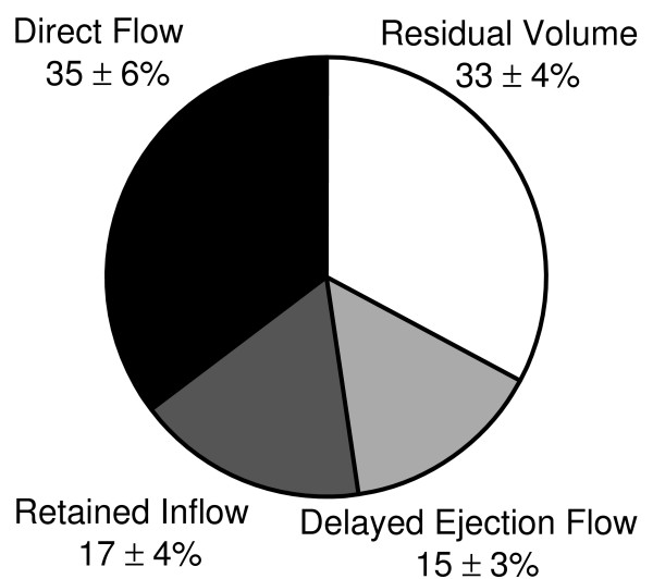
The volume and distribution of the LV flow components were determined in all subjects. The calculated LV outflow volumes [ml] (67 +/- 13) appeared to fall in between those obtained by through-plane phase-contrast CMR (77 +/- 16) and Doppler ultrasound (58 +/- 10), respectively. Calculated volumes of LV inflow (68 +/- 11) and outflow (67 +/- 13) were well matched (NS). Low inter- and intra-observer variability for the assessment of the volumes of the flow components was obtained.

CONCLUSIONS

This semi-automatic analysis approach for the quantification of 4D blood flow resulted in accurate LV inflow and outflow volumes and a high reproducibility for the assessment of LV flow components.

摘要

背景

跳动的心脏是心血管系统中血流的发生器。在心脏自身的腔室内,正常的复杂血流模式可能会被疾病扰乱。缺乏用于量化具有 4D(3D+时间)性质的心脏内血流的方法。我们试图开发和验证一种新的半自动分析方法,该方法集成了流量和形态数据。

方法

在六名健康受试者和三名扩张型心肌病患者中,采集了三个方向的三维电影相位对比心血管磁共振(CMR)速度数据和平衡稳态自由进动长轴和短轴图像。在等容收缩(IVC)和等容舒张(IVR)时,从短轴图像上分割 LV 心内膜。在 IVC 时,从 IVC LV 血容量发出轨迹线,并在时间上向前和向后追踪,直到 IVR,从而包括整个心动周期。使用 IVR 体积来确定轨迹线是否以及在何处离开 LV。此信息用于自动将轨迹线分为四种不同的流动成分:直接流动、保留流入、延迟射流和残留体积。通过将轨迹线的数量乘以每条轨迹线所代表的血容量,计算每个成分的血容量。通过分析 LV 流入和流出量、四个流动成分以及舒张末期容积,评估该方法的准确性和观察者内和观察者间的可重复性。

结果

在所有受试者中确定了 LV 流动成分的体积和分布。计算出的 LV 流出量[ml](67 +/- 13)似乎介于平面相位对比 CMR(77 +/- 16)和多普勒超声(58 +/- 10)获得的值之间。计算出的 LV 流入量(68 +/- 11)和流出量(67 +/- 13)非常匹配(NS)。评估流量成分体积的观察者内和观察者间的变异性较低。

结论

这种用于量化 4D 血流的半自动分析方法可准确测量 LV 流入和流出量,并高度重现性地评估 LV 流量成分。

https://cdn.ncbi.nlm.nih.gov/pmc/blobs/3d2c/2831022/e2b904c1abab/1532-429X-12-9-4.jpg
https://cdn.ncbi.nlm.nih.gov/pmc/blobs/3d2c/2831022/451b31a6c382/1532-429X-12-9-1.jpg
https://cdn.ncbi.nlm.nih.gov/pmc/blobs/3d2c/2831022/ce9d976c5b9f/1532-429X-12-9-2.jpg
https://cdn.ncbi.nlm.nih.gov/pmc/blobs/3d2c/2831022/9a6171ed6136/1532-429X-12-9-3.jpg
https://cdn.ncbi.nlm.nih.gov/pmc/blobs/3d2c/2831022/e2b904c1abab/1532-429X-12-9-4.jpg

相似文献

1
Semi-automatic quantification of 4D left ventricular blood flow.四维左心室血流半自动定量。
J Cardiovasc Magn Reson. 2010 Feb 12;12(1):9. doi: 10.1186/1532-429X-12-9.
2
Test-retest variability of left ventricular 4D flow cardiovascular magnetic resonance measurements in healthy subjects.健康受试者左心室 4D 血流心血管磁共振测量的重测变异性。
J Cardiovasc Magn Reson. 2018 Mar 2;20(1):15. doi: 10.1186/s12968-018-0432-4.
3
Diastolic function assessment with four-dimensional flow cardiovascular magnetic resonance using automatic deep learning E/A ratio analysis.四维血流心血管磁共振自动深度学习法评估舒张功能。
J Cardiovasc Magn Reson. 2024 Summer;26(1):101042. doi: 10.1016/j.jocmr.2024.101042. Epub 2024 Mar 30.
4
Compressed sensing real-time cine cardiovascular magnetic resonance: accurate assessment of left ventricular function in a single-breath-hold.压缩感知实时电影心血管磁共振成像:单次屏气下对左心室功能的准确评估
J Cardiovasc Magn Reson. 2016 Aug 24;18(1):50. doi: 10.1186/s12968-016-0271-0.
5
Deep learning based automated left ventricle segmentation and flow quantification in 4D flow cardiac MRI.基于深度学习的 4D 流心脏 MRI 中左心室自动分割和流量定量
J Cardiovasc Magn Reson. 2024 Summer;26(1):100003. doi: 10.1016/j.jocmr.2023.100003. Epub 2024 Jan 10.
6
Comparison of different methods for the estimation of aortic pulse wave velocity from 4D flow cardiovascular magnetic resonance.从 4D 流心血管磁共振评估主动脉脉搏波速度的不同方法比较。
J Cardiovasc Magn Reson. 2019 Dec 12;21(1):75. doi: 10.1186/s12968-019-0584-x.
7
Atlas-based analysis of 4D flow CMR: automated vessel segmentation and flow quantification.基于图谱的4D流心脏磁共振成像分析:自动血管分割与血流定量
J Cardiovasc Magn Reson. 2015 Oct 5;17:87. doi: 10.1186/s12968-015-0190-5.
8
Automated left ventricular diastolic function evaluation from phase-contrast cardiovascular magnetic resonance and comparison with Doppler echocardiography.基于相位对比心血管磁共振的左心室舒张功能自动评估,并与多普勒超声心动图比较。
J Cardiovasc Magn Reson. 2010 Nov 9;12(1):63. doi: 10.1186/1532-429X-12-63.
9
Comparison of long and short axis quantification of left ventricular volume parameters by cardiovascular magnetic resonance, with ex-vivo validation.心血管磁共振长轴和短轴左心室容积参数定量比较及其与离体验证。
J Cardiovasc Magn Reson. 2011 Aug 11;13(1):40. doi: 10.1186/1532-429X-13-40.
10
Mechanical dyssynchrony alters left ventricular flow energetics in failing hearts with LBBB: a 4D flow CMR pilot study.机械性不同步改变左束支传导阻滞所致心力衰竭患者左心室血流能量学:一项四维血流心脏磁共振成像初步研究。
Int J Cardiovasc Imaging. 2018 Apr;34(4):587-596. doi: 10.1007/s10554-017-1261-5. Epub 2017 Nov 2.

引用本文的文献

1
Flow transport and not ejection fraction determines blood stasis in patients with impaired left ventricular systolic function.在左心室收缩功能受损的患者中,血流运输而非射血分数决定了血液瘀滞。
Physiol Rep. 2025 Jul;13(13):e70351. doi: 10.14814/phy2.70351.
2
The effect of respiratory motion compensation in intracardiac 4D flow magnetic resonance imaging on left ventricular flow dynamics, multicomponent particle tracing, and valve tracking.心脏内4D血流磁共振成像中呼吸运动补偿对左心室血流动力学、多组分粒子追踪和瓣膜追踪的影响。
Eur Heart J Imaging Methods Pract. 2025 Mar 10;3(1):qyaf020. doi: 10.1093/ehjimp/qyaf020. eCollection 2025 Jan.
3

本文引用的文献

1
Fluid-dynamic modeling of the human left ventricle: methodology and application to surgical ventricular reconstruction.人体左心室的流体动力学建模:方法及其在外科心室重建中的应用
Ann Thorac Surg. 2009 Apr;87(4):1187-95. doi: 10.1016/j.athoracsur.2009.01.036.
2
Quantitative 2D and 3D phase contrast MRI: optimized analysis of blood flow and vessel wall parameters.定量二维和三维相位对比磁共振成像:血流和血管壁参数的优化分析
Magn Reson Med. 2008 Nov;60(5):1218-31. doi: 10.1002/mrm.21778.
3
Assessment of fluctuating velocities in disturbed cardiovascular blood flow: in vivo feasibility of generalized phase-contrast MRI.
Understanding Right Heart Flow: Implications for Interatrial Shunt Device Therapy in Heart Failure.
了解右心血流:对心力衰竭患者房间隔分流装置治疗的意义。
J Soc Cardiovasc Angiogr Interv. 2025 Jan 21;4(1):102439. doi: 10.1016/j.jscai.2024.102439. eCollection 2025 Jan.
4
Right Ventricular Dysfunction in Cardiac Anesthesia: Perioperative Assessment and Underlying Mechanisms.心脏麻醉中的右心室功能障碍:围手术期评估及潜在机制
Rev Cardiovasc Med. 2025 Feb 12;26(2):26286. doi: 10.31083/RCM26286. eCollection 2025 Feb.
5
Characterization of Intracardiac Flow in the Right Ventricle With Pressure and Volume Overload in Children.儿童右心室压力和容量超负荷时心内血流的特征分析
Cardiol Res. 2025 Feb;16(1):22-32. doi: 10.14740/cr2009. Epub 2025 Jan 6.
6
Intracardiac fluid dynamic analysis: available techniques and novel clinical applications.心内流体动力学分析:现有技术及新的临床应用
BMC Cardiovasc Disord. 2024 Dec 19;24(1):716. doi: 10.1186/s12872-024-04371-3.
7
Super-resolution left ventricular flow and pressure mapping by Navier-Stokes-informed neural networks.基于纳维-斯托克斯方程的神经网络实现超分辨率左心室血流和压力映射
Comput Biol Med. 2025 Feb;185:109476. doi: 10.1016/j.compbiomed.2024.109476. Epub 2024 Dec 12.
8
Super-resolution Left Ventricular Flow and Pressure Mapping by Navier-Stokes-Informed Neural Networks.基于纳维-斯托克斯信息神经网络的超分辨率左心室血流和压力映射
bioRxiv. 2024 Apr 15:2024.04.12.589319. doi: 10.1101/2024.04.12.589319.
9
Deep learning based automated left ventricle segmentation and flow quantification in 4D flow cardiac MRI.基于深度学习的 4D 流心脏 MRI 中左心室自动分割和流量定量
J Cardiovasc Magn Reson. 2024 Summer;26(1):100003. doi: 10.1016/j.jocmr.2023.100003. Epub 2024 Jan 10.
10
Comprehensive left atrial flow component analysis reveals abnormal flow patterns in paroxysmal atrial fibrillation.全面的左心房流分量分析揭示阵发性心房颤动中的异常血流模式。
Am J Physiol Heart Circ Physiol. 2024 Mar 1;326(3):H511-H521. doi: 10.1152/ajpheart.00614.2023. Epub 2023 Dec 22.
评估心血管紊乱血流中的波动速度:广义相位对比磁共振成像的体内可行性。
J Magn Reson Imaging. 2008 Sep;28(3):655-63. doi: 10.1002/jmri.21475.
4
The looped heart does not save energy by maintaining the momentum of blood flowing in the ventricle.环状心脏并不会通过维持心室中血液流动的动量来节省能量。
Am J Physiol Heart Circ Physiol. 2008 May;294(5):H2191-6. doi: 10.1152/ajpheart.00041.2008. Epub 2008 Mar 7.
5
Comparison of flow patterns in ascending aortic aneurysms and volunteers using four-dimensional magnetic resonance velocity mapping.使用四维磁共振速度成像技术比较升主动脉瘤患者与志愿者的血流模式。
J Magn Reson Imaging. 2007 Dec;26(6):1471-9. doi: 10.1002/jmri.21082.
6
Transit of blood flow through the human left ventricle mapped by cardiovascular magnetic resonance.通过心血管磁共振成像绘制的血液流经人体左心室的过程。
J Cardiovasc Magn Reson. 2007;9(5):741-7. doi: 10.1080/10976640701544530.
7
Cardiology is flow.心脏病学就是血流。
Circulation. 2006 Jun 13;113(23):2679-82. doi: 10.1161/CIRCULATIONAHA.106.632687.
8
Effect of endocardial trabeculae on left ventricular measurements and measurement reproducibility at cardiovascular MR imaging.心内膜小梁对心血管磁共振成像中左心室测量及测量可重复性的影响。
Radiology. 2005 Jul;236(1):57-64. doi: 10.1148/radiol.2353040601. Epub 2005 Jun 13.
9
Time-resolved 3-dimensional velocity mapping in the thoracic aorta: visualization of 3-directional blood flow patterns in healthy volunteers and patients.胸主动脉的时间分辨三维速度映射:健康志愿者和患者三维血流模式的可视化
J Comput Assist Tomogr. 2004 Jul-Aug;28(4):459-68. doi: 10.1097/00004728-200407000-00005.
10
Flow patterns in the aortic root and the aorta studied with time-resolved, 3-dimensional, phase-contrast magnetic resonance imaging: implications for aortic valve-sparing surgery.采用时间分辨三维相位对比磁共振成像研究主动脉根部和主动脉内的血流模式:对保留主动脉瓣手术的意义
J Thorac Cardiovasc Surg. 2004 Jun;127(6):1602-7. doi: 10.1016/j.jtcvs.2003.10.042.