• 文献检索
  • 文档翻译
  • 深度研究
  • 学术资讯
  • Suppr Zotero 插件Zotero 插件
  • 邀请有礼
  • 套餐&价格
  • 历史记录
应用&插件
Suppr Zotero 插件Zotero 插件浏览器插件Mac 客户端Windows 客户端微信小程序
定价
高级版会员购买积分包购买API积分包
服务
文献检索文档翻译深度研究API 文档MCP 服务
关于我们
关于 Suppr公司介绍联系我们用户协议隐私条款
关注我们

Suppr 超能文献

核心技术专利:CN118964589B侵权必究
粤ICP备2023148730 号-1Suppr @ 2026

文献检索

告别复杂PubMed语法,用中文像聊天一样搜索,搜遍4000万医学文献。AI智能推荐,让科研检索更轻松。

立即免费搜索

文件翻译

保留排版,准确专业,支持PDF/Word/PPT等文件格式,支持 12+语言互译。

免费翻译文档

深度研究

AI帮你快速写综述,25分钟生成高质量综述,智能提取关键信息,辅助科研写作。

立即免费体验

分割组织病理学图像:一种用于监督的颜色-纹理分割和细胞分裂的集成框架。

Partitioning histopathological images: an integrated framework for supervised color-texture segmentation and cell splitting.

机构信息

Department of Biomedical Informatics, Ohio State University, Columbus, OH 43201, USA.

出版信息

IEEE Trans Med Imaging. 2011 Sep;30(9):1661-77. doi: 10.1109/TMI.2011.2141674. Epub 2011 Apr 11.

DOI:10.1109/TMI.2011.2141674
PMID:21486712
原文链接:https://pmc.ncbi.nlm.nih.gov/articles/PMC3165069/
Abstract

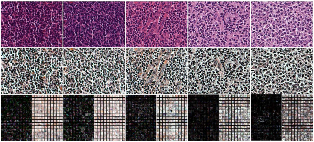
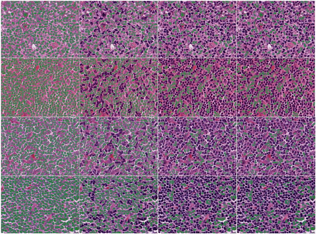
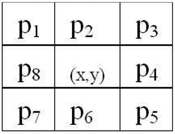
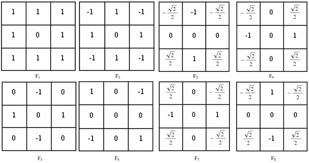
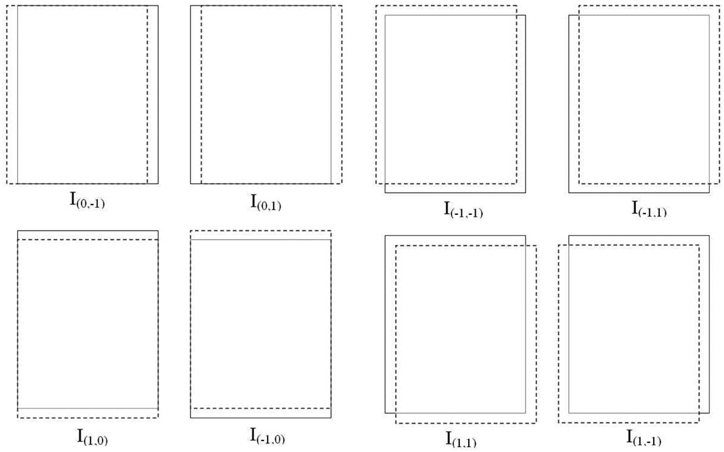
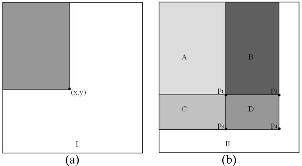
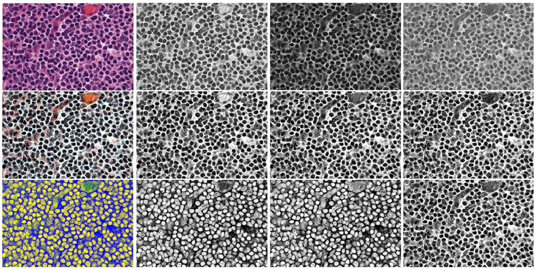
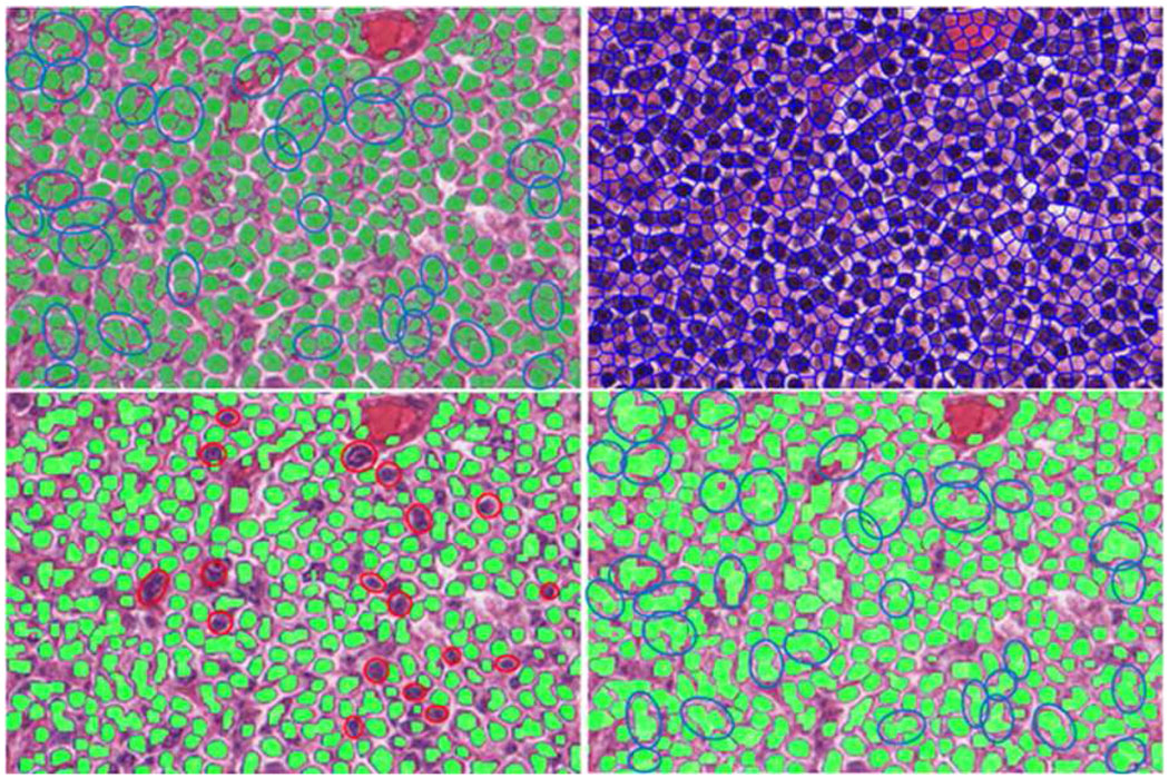
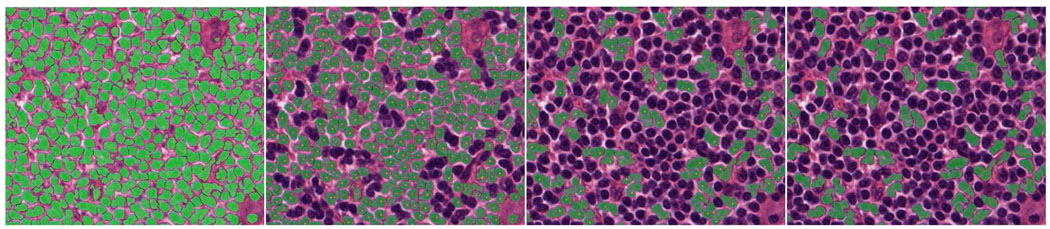
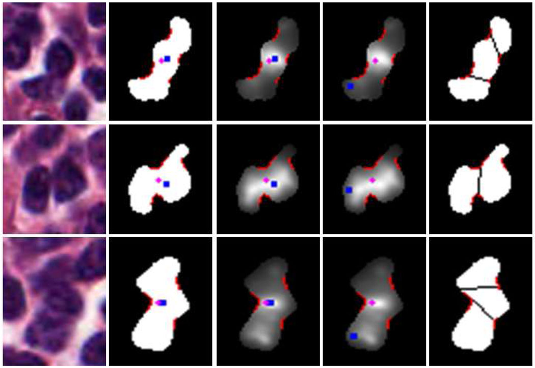
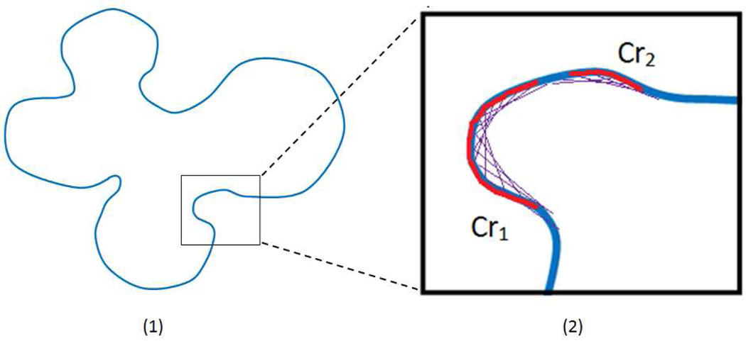
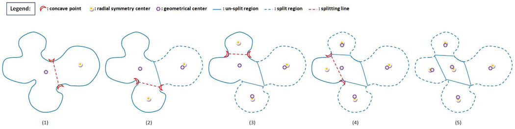
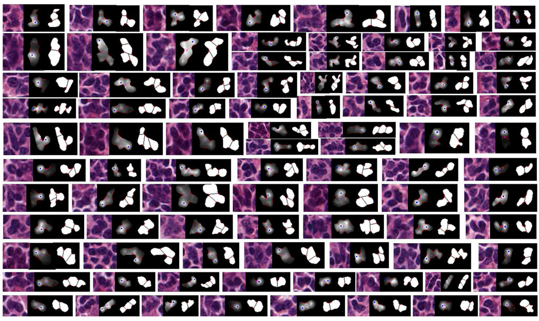
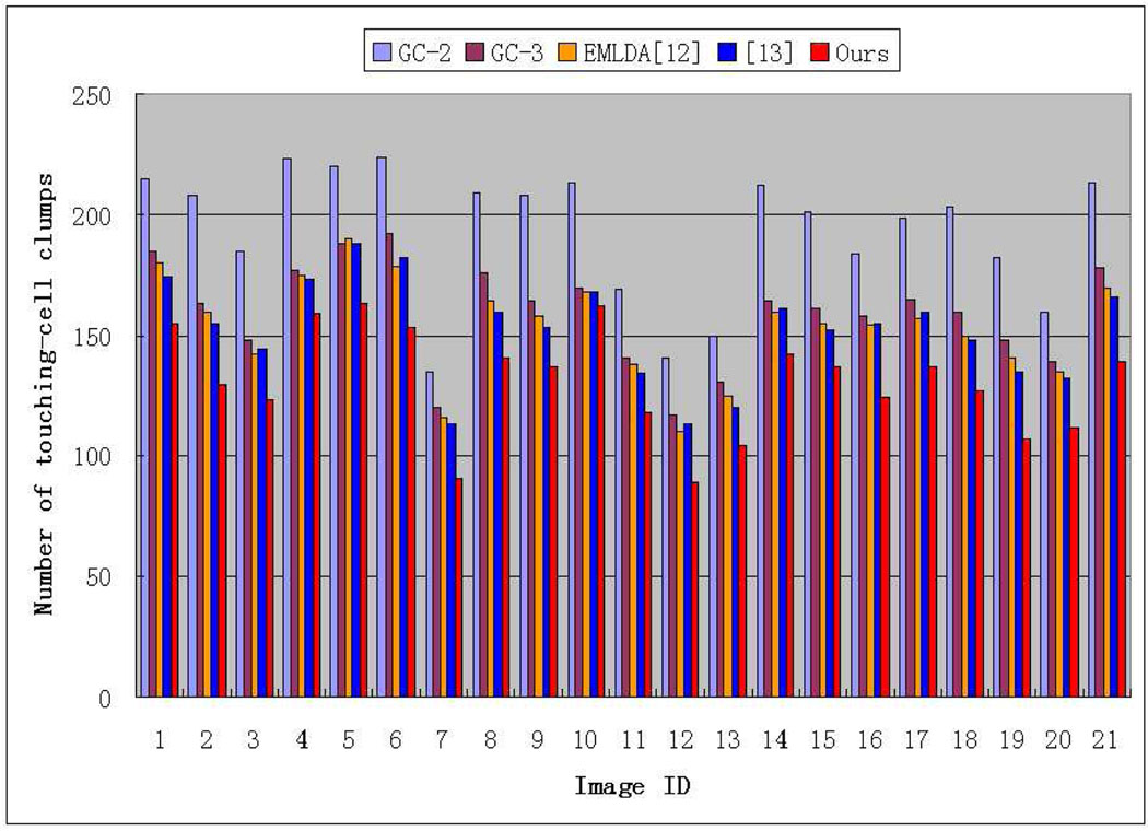
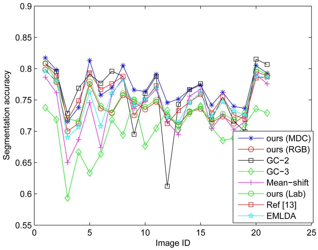
For quantitative analysis of histopathological images, such as the lymphoma grading systems, quantification of features is usually carried out on single cells before categorizing them by classification algorithms. To this end, we propose an integrated framework consisting of a novel supervised cell-image segmentation algorithm and a new touching-cell splitting method. For the segmentation part, we segment the cell regions from the other areas by classifying the image pixels into either cell or extra-cellular category. Instead of using pixel color intensities, the color-texture extracted at the local neighborhood of each pixel is utilized as the input to our classification algorithm. The color-texture at each pixel is extracted by local Fourier transform (LFT) from a new color space, the most discriminant color space (MDC). The MDC color space is optimized to be a linear combination of the original RGB color space so that the extracted LFT texture features in the MDC color space can achieve most discrimination in terms of classification (segmentation) performance. To speed up the texture feature extraction process, we develop an efficient LFT extraction algorithm based on image shifting and image integral. For the splitting part, given a connected component of the segmentation map, we initially differentiate whether it is a touching-cell clump or a single nontouching cell. The differentiation is mainly based on the distance between the most likely radial-symmetry center and the geometrical center of the connected component. The boundaries of touching-cell clumps are smoothed out by Fourier shape descriptor before carrying out an iterative, concave-point and radial-symmetry based splitting algorithm. To test the validity, effectiveness and efficiency of the framework, it is applied to follicular lymphoma pathological images, which exhibit complex background and extracellular texture with nonuniform illumination condition. For comparison purposes, the results of the proposed segmentation algorithm are evaluated against the outputs of superpixel, graph-cut, mean-shift, and two state-of-the-art pathological image segmentation methods using ground-truth that was established by manual segmentation of cells in the original images. Our segmentation algorithm achieves better results than the other compared methods. The results of splitting are evaluated in terms of under-splitting, over-splitting, and encroachment errors. By summing up the three types of errors, we achieve a total error rate of 5.25% per image.

摘要

对于组织病理学图像的定量分析,例如淋巴瘤分级系统,通常在通过分类算法对其进行分类之前,对单个细胞的特征进行量化。为此,我们提出了一个由新的有监督细胞图像分割算法和新的粘连细胞分割方法组成的集成框架。对于分割部分,我们通过将图像像素分类为细胞或细胞外类别,将细胞区域与其他区域分开。我们的分类算法不使用像素颜色强度,而是利用每个像素的局部邻域提取的颜色纹理作为输入。颜色纹理是通过局部傅里叶变换(LFT)从新的颜色空间(最具判别力的颜色空间,MDC)中提取的。MDC 颜色空间被优化为原始 RGB 颜色空间的线性组合,以使 MDC 颜色空间中提取的 LFT 纹理特征在分类(分割)性能方面具有最大的可区分性。为了加速纹理特征提取过程,我们开发了一种基于图像移位和图像积分的高效 LFT 提取算法。对于分割部分,给定分割图的一个连通分量,我们最初区分它是粘连细胞团还是单个非粘连细胞。这种区分主要基于最可能的径向对称中心与连通分量的几何中心之间的距离。在进行迭代、凹点和基于径向对称的分割算法之前,先通过傅里叶形状描述符对粘连细胞团的边界进行平滑处理。为了测试框架的有效性、有效性和效率,将其应用于滤泡性淋巴瘤病理图像,这些图像表现出复杂的背景和具有不均匀照明条件的细胞外纹理。为了进行比较,使用原始图像中手动分割细胞建立的真实分割,评估了所提出的分割算法与超像素、图割、均值漂移和两种最先进的病理图像分割方法的输出结果。我们的分割算法比其他比较方法取得了更好的结果。分割结果根据欠分割、过分割和入侵误差进行评估。通过总结这三种类型的误差,我们获得了每张图像 5.25%的总误差率。

https://cdn.ncbi.nlm.nih.gov/pmc/blobs/8a2a/3165069/ab0c3d0b7a44/nihms299955f16.jpg
https://cdn.ncbi.nlm.nih.gov/pmc/blobs/8a2a/3165069/fcd01908056b/nihms299955f1.jpg
https://cdn.ncbi.nlm.nih.gov/pmc/blobs/8a2a/3165069/77f38e16c3ec/nihms299955f2.jpg
https://cdn.ncbi.nlm.nih.gov/pmc/blobs/8a2a/3165069/fd1b035da410/nihms299955f3.jpg
https://cdn.ncbi.nlm.nih.gov/pmc/blobs/8a2a/3165069/aeb37aa9f817/nihms299955f4.jpg
https://cdn.ncbi.nlm.nih.gov/pmc/blobs/8a2a/3165069/890767173849/nihms299955f5.jpg
https://cdn.ncbi.nlm.nih.gov/pmc/blobs/8a2a/3165069/758278571597/nihms299955f6.jpg
https://cdn.ncbi.nlm.nih.gov/pmc/blobs/8a2a/3165069/eedf7f2a13c2/nihms299955f7.jpg
https://cdn.ncbi.nlm.nih.gov/pmc/blobs/8a2a/3165069/8baec12ee4ce/nihms299955f8.jpg
https://cdn.ncbi.nlm.nih.gov/pmc/blobs/8a2a/3165069/03dd25413adb/nihms299955f9.jpg
https://cdn.ncbi.nlm.nih.gov/pmc/blobs/8a2a/3165069/f59c769d89e5/nihms299955f10.jpg
https://cdn.ncbi.nlm.nih.gov/pmc/blobs/8a2a/3165069/99f4274a76fe/nihms299955f11.jpg
https://cdn.ncbi.nlm.nih.gov/pmc/blobs/8a2a/3165069/93cf6017267b/nihms299955f12.jpg
https://cdn.ncbi.nlm.nih.gov/pmc/blobs/8a2a/3165069/e82ed985f9be/nihms299955f13.jpg
https://cdn.ncbi.nlm.nih.gov/pmc/blobs/8a2a/3165069/8c5dc63642b3/nihms299955f14.jpg
https://cdn.ncbi.nlm.nih.gov/pmc/blobs/8a2a/3165069/9e33b02f3c2e/nihms299955f15.jpg
https://cdn.ncbi.nlm.nih.gov/pmc/blobs/8a2a/3165069/ab0c3d0b7a44/nihms299955f16.jpg

相似文献

1
Partitioning histopathological images: an integrated framework for supervised color-texture segmentation and cell splitting.分割组织病理学图像:一种用于监督的颜色-纹理分割和细胞分裂的集成框架。
IEEE Trans Med Imaging. 2011 Sep;30(9):1661-77. doi: 10.1109/TMI.2011.2141674. Epub 2011 Apr 11.
2
CTex--an adaptive unsupervised segmentation algorithm based on color-texture coherence.CTex——一种基于颜色纹理一致性的自适应无监督分割算法。
IEEE Trans Image Process. 2008 Oct;17(10):1926-39. doi: 10.1109/TIP.2008.2001047.
3
Supervised learning-based cell image segmentation for p53 immunohistochemistry.基于监督学习的p53免疫组织化学细胞图像分割
IEEE Trans Biomed Eng. 2006 Jun;53(6):1153-63. doi: 10.1109/TBME.2006.873538.
4
Automatic image segmentation by dynamic region growth and multiresolution merging.通过动态区域生长和多分辨率合并实现自动图像分割。
IEEE Trans Image Process. 2009 Oct;18(10):2275-88. doi: 10.1109/TIP.2009.2025555. Epub 2009 Jun 16.
5
Color image segmentation based on mean shift and normalized cuts.基于均值漂移和归一化切割的彩色图像分割
IEEE Trans Syst Man Cybern B Cybern. 2007 Oct;37(5):1382-9. doi: 10.1109/tsmcb.2007.902249.
6
Graph run-length matrices for histopathological image segmentation.用于组织病理学图像分割的图游程长度矩阵。
IEEE Trans Med Imaging. 2011 Mar;30(3):721-32. doi: 10.1109/TMI.2010.2094200. Epub 2010 Nov 22.
7
Charisma: an integrated approach to automatic H&E-stained skeletal muscle cell segmentation using supervised learning and novel robust clump splitting.魅力:一种基于监督学习和新型稳健聚类分割的自动 H&E 染色骨骼肌细胞分割的综合方法。
Med Image Anal. 2013 Dec;17(8):1206-19. doi: 10.1016/j.media.2013.07.007. Epub 2013 Aug 13.
8
A wrapper-based approach to image segmentation and classification.一种基于包装器的图像分割与分类方法。
IEEE Trans Image Process. 2005 Dec;14(12):2060-72. doi: 10.1109/tip.2005.859374.
9
Segmentation of psoriasis vulgaris images using multiresolution-based orthogonal subspace techniques.基于多分辨率的正交子空间技术对寻常型银屑病图像进行分割。
IEEE Trans Syst Man Cybern B Cybern. 2006 Apr;36(2):390-402. doi: 10.1109/tsmcb.2005.859935.
10
Image segmentation based on GrabCut framework integrating multiscale nonlinear structure tensor.基于集成多尺度非线性结构张量的GrabCut框架的图像分割
IEEE Trans Image Process. 2009 Oct;18(10):2289-302. doi: 10.1109/TIP.2009.2025560. Epub 2009 Jun 16.

引用本文的文献

1
A Novel Heteromorphous Convolutional Neural Network for Automated Assessment of Tumors in Colon and Lung Histopathology Images.一种用于结肠和肺部组织病理学图像中肿瘤自动评估的新型异质卷积神经网络。
Biomimetics (Basel). 2023 Aug 16;8(4):370. doi: 10.3390/biomimetics8040370.
2
3D reconstruction of skin and spatial mapping of immune cell density, vascular distance and effects of sun exposure and aging.皮肤的 3D 重建和免疫细胞密度、血管距离的空间映射,以及日光照射和衰老的影响。
Commun Biol. 2023 Jul 19;6(1):718. doi: 10.1038/s42003-023-04991-z.
3
Cervical cell nuclei segmentation based on GC-UNet.基于GC-UNet的宫颈细胞核分割
Heliyon. 2023 Jun 28;9(7):e17647. doi: 10.1016/j.heliyon.2023.e17647. eCollection 2023 Jul.
4
ML-Based Texture and Wavelet Features Extraction Technique to Predict Gastric Mesothelioma Cancer.基于机器学习的纹理和小波特征提取技术预测胃间皮瘤癌症。
Biomed Res Int. 2022 Jul 4;2022:1012684. doi: 10.1155/2022/1012684. eCollection 2022.
5
Synthetic-to-real: instance segmentation of clinical cluster cells with unlabeled synthetic training.合成到真实:使用未标记的合成训练进行临床聚类细胞的实例分割。
Bioinformatics. 2022 Jun 24;38(Suppl 1):i53-i59. doi: 10.1093/bioinformatics/btac219.
6
Content-Based Image Retrieval Using Colour, Gray, Advanced Texture, Shape Features, and Random Forest Classifier with Optimized Particle Swarm Optimization.基于内容的图像检索:结合颜色、灰度、高级纹理、形状特征以及采用优化粒子群优化的随机森林分类器
Int J Biomed Imaging. 2022 Apr 21;2022:3211793. doi: 10.1155/2022/3211793. eCollection 2022.
7
Graph-Theoretic Post-Processing of Segmentation With Application to Dense Biofilms.基于图论的分割后处理及其在密集生物膜中的应用。
IEEE Trans Image Process. 2021;30:8580-8594. doi: 10.1109/TIP.2021.3116792. Epub 2021 Oct 15.
8
Detection of Cervical Cancer Cells in Whole Slide Images Using Deformable and Global Context Aware Faster RCNN-FPN.使用可变形和全局上下文感知的 Faster RCNN-FPN 检测全幻灯片图像中的宫颈癌细胞。
Curr Oncol. 2021 Sep 16;28(5):3585-3601. doi: 10.3390/curroncol28050307.
9
Accurate Segmentation of Nuclear Regions with Multi-Organ Histopathology Images Using Artificial Intelligence for Cancer Diagnosis in Personalized Medicine.利用人工智能对多器官组织病理学图像中的核区域进行精确分割,用于个性化医疗中的癌症诊断。
J Pers Med. 2021 Jun 4;11(6):515. doi: 10.3390/jpm11060515.
10
An automatic nuclei segmentation method based on deep convolutional neural networks for histopathology images.一种基于深度卷积神经网络的组织病理学图像细胞核自动分割方法。
BMC Biomed Eng. 2019 Oct 17;1:24. doi: 10.1186/s42490-019-0026-8. eCollection 2019.

本文引用的文献

1
Computer-aided evaluation of neuroblastoma on whole-slide histology images: Classifying grade of neuroblastic differentiation.基于全切片组织学图像的神经母细胞瘤计算机辅助评估:神经母细胞分化程度分类
Pattern Recognit. 2009 Jun;42(6):1080-1092. doi: 10.1016/j.patcog.2008.10.035.
2
Histopathological image analysis: a review.组织病理学图像分析:综述。
IEEE Rev Biomed Eng. 2009;2:147-71. doi: 10.1109/RBME.2009.2034865. Epub 2009 Oct 30.
3
Computer-aided detection of centroblasts for follicular lymphoma grading using adaptive likelihood-based cell segmentation.基于自适应似然的细胞分割的滤泡性淋巴瘤分级中中心母细胞的计算机辅助检测。
IEEE Trans Biomed Eng. 2010 Oct;57(10):2613-6. doi: 10.1109/TBME.2010.2055058. Epub 2010 Jun 28.
4
Expectation-maximization-driven geodesic active contour with overlap resolution (EMaGACOR): application to lymphocyte segmentation on breast cancer histopathology.基于期望最大化的带重叠解析的测地线主动轮廓(EMaGACOR):在乳腺癌组织病理学淋巴细胞分割中的应用。
IEEE Trans Biomed Eng. 2010 Jul;57(7):1676-89. doi: 10.1109/TBME.2010.2041232. Epub 2010 Feb 17.
5
Improved automatic detection and segmentation of cell nuclei in histopathology images.改进组织病理学图像中细胞核的自动检测和分割。
IEEE Trans Biomed Eng. 2010 Apr;57(4):841-52. doi: 10.1109/TBME.2009.2035102. Epub 2009 Oct 30.
6
Segmentation of clustered nuclei with shape markers and marking function.利用形状标记和标记函数对聚集细胞核进行分割。
IEEE Trans Biomed Eng. 2009 Mar;56(3):741-8. doi: 10.1109/TBME.2008.2008635. Epub 2008 Nov 11.
7
Adaptive discriminant wavelet packet transform and local binary patterns for meningioma subtype classification.用于脑膜瘤亚型分类的自适应判别小波包变换与局部二值模式
Med Image Comput Comput Assist Interv. 2008;11(Pt 2):196-204. doi: 10.1007/978-3-540-85990-1_24.
8
Automatic image analysis of histopathology specimens using concave vertex graph.使用凹顶点图对组织病理学标本进行自动图像分析。
Med Image Comput Comput Assist Interv. 2008;11(Pt 1):833-41. doi: 10.1007/978-3-540-85988-8_99.
9
Segmenting and counting of wall-pasted cells based on gabor filter.
Conf Proc IEEE Eng Med Biol Soc. 2005;2005:3324-7. doi: 10.1109/IEMBS.2005.1617188.
10
Supervised learning-based cell image segmentation for p53 immunohistochemistry.基于监督学习的p53免疫组织化学细胞图像分割
IEEE Trans Biomed Eng. 2006 Jun;53(6):1153-63. doi: 10.1109/TBME.2006.873538.