• 文献检索
  • 文档翻译
  • 深度研究
  • 学术资讯
  • Suppr Zotero 插件Zotero 插件
  • 邀请有礼
  • 套餐&价格
  • 历史记录
应用&插件
Suppr Zotero 插件Zotero 插件浏览器插件Mac 客户端Windows 客户端微信小程序
定价
高级版会员购买积分包购买API积分包
服务
文献检索文档翻译深度研究API 文档MCP 服务
关于我们
关于 Suppr公司介绍联系我们用户协议隐私条款
关注我们

Suppr 超能文献

核心技术专利:CN118964589B侵权必究
粤ICP备2023148730 号-1Suppr @ 2026

文献检索

告别复杂PubMed语法,用中文像聊天一样搜索,搜遍4000万医学文献。AI智能推荐,让科研检索更轻松。

立即免费搜索

文件翻译

保留排版,准确专业,支持PDF/Word/PPT等文件格式,支持 12+语言互译。

免费翻译文档

深度研究

AI帮你快速写综述,25分钟生成高质量综述,智能提取关键信息,辅助科研写作。

立即免费体验

基于全切片组织学图像的神经母细胞瘤计算机辅助评估:神经母细胞分化程度分类

Computer-aided evaluation of neuroblastoma on whole-slide histology images: Classifying grade of neuroblastic differentiation.

作者信息

Kong J, Sertel O, Shimada H, Boyer K L, Saltz J H, Gurcan M N

机构信息

Department of Electrical and Computer Engineering, The Ohio State University, 2015 Neil Avenue, Columbus, OH 43210, USA.

Department of Biomedical Informatics, The Ohio State University, 3190 Graves Hall, Columbus, OH 43210, USA.

出版信息

Pattern Recognit. 2009 Jun;42(6):1080-1092. doi: 10.1016/j.patcog.2008.10.035.

DOI:10.1016/j.patcog.2008.10.035
PMID:28626265
原文链接:https://pmc.ncbi.nlm.nih.gov/articles/PMC5473636/
Abstract

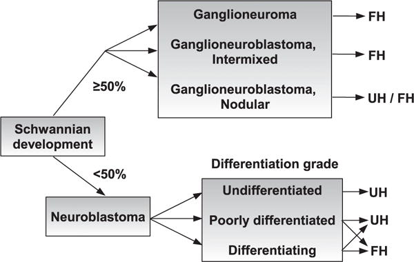
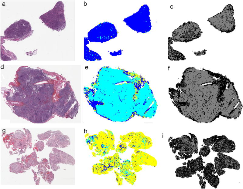
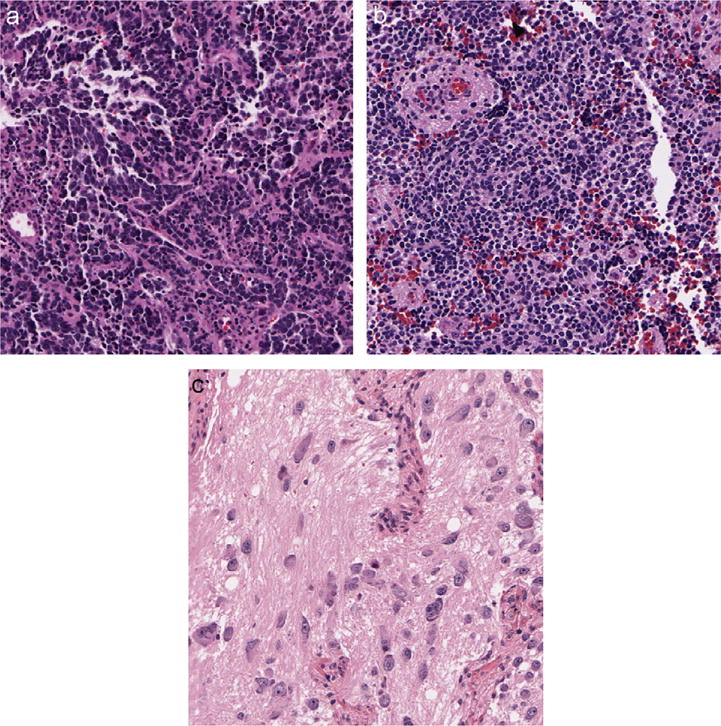
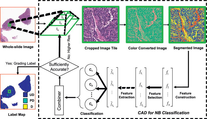
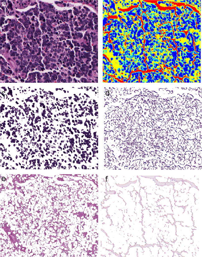
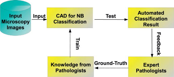
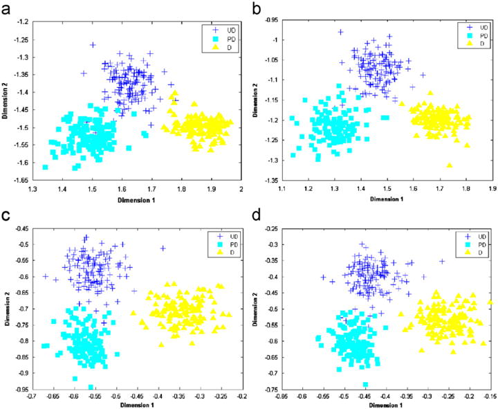
Neuroblastoma (NB) is one of the most frequently occurring cancerous tumors in children. The current grading evaluations for patients with this disease require pathologists to identify certain morphological characteristics with microscopic examinations of tumor tissues. Thanks to the advent of modern digital scanners, it is now feasible to scan cross-section tissue specimens and acquire whole-slide digital images. As a result, computerized analysis of these images can generate key quantifiable parameters and assist pathologists with grading evaluations. In this study, image analysis techniques are applied to histological images of haematoxylin and eosin (H&E) stained slides for identifying image regions associated with different pathological components. Texture features derived from segmented components of tissues are extracted and processed by an automated classifier group trained with sample images with different grades of neuroblastic differentiation in a multi-resolution framework. The trained classification system is tested on 33 whole-slide tumor images. The resulting whole-slide classification accuracy produced by the computerized system is 87.88%. Therefore, the developed system is a promising tool to facilitate grading whole-slide images of NB biopsies with high throughput.

摘要

神经母细胞瘤(NB)是儿童中最常见的癌性肿瘤之一。目前针对该疾病患者的分级评估需要病理学家通过对肿瘤组织进行显微镜检查来识别某些形态学特征。得益于现代数字扫描仪的出现,现在扫描横截面组织标本并获取全切片数字图像成为可能。因此,对这些图像进行计算机分析可以生成关键的可量化参数,并协助病理学家进行分级评估。在本研究中,图像分析技术被应用于苏木精和伊红(H&E)染色玻片的组织学图像,以识别与不同病理成分相关的图像区域。从组织的分割成分中提取纹理特征,并在多分辨率框架中由使用具有不同神经母细胞分化等级的样本图像训练的自动分类器组进行处理。训练好的分类系统在33张全切片肿瘤图像上进行测试。计算机系统产生的全切片分类准确率为87.88%。因此,所开发的系统是一种很有前景的工具,可用于高通量地对NB活检的全切片图像进行分级。

https://cdn.ncbi.nlm.nih.gov/pmc/blobs/0488/5473636/7022bc152bb7/nihms867505f9.jpg
https://cdn.ncbi.nlm.nih.gov/pmc/blobs/0488/5473636/2111dfafda98/nihms867505f1.jpg
https://cdn.ncbi.nlm.nih.gov/pmc/blobs/0488/5473636/111b86b427f0/nihms867505f2.jpg
https://cdn.ncbi.nlm.nih.gov/pmc/blobs/0488/5473636/df7f35be69a0/nihms867505f3.jpg
https://cdn.ncbi.nlm.nih.gov/pmc/blobs/0488/5473636/ca0c0897cf1a/nihms867505f4.jpg
https://cdn.ncbi.nlm.nih.gov/pmc/blobs/0488/5473636/0225a862441c/nihms867505f5.jpg
https://cdn.ncbi.nlm.nih.gov/pmc/blobs/0488/5473636/bd353a2ebb7c/nihms867505f6.jpg
https://cdn.ncbi.nlm.nih.gov/pmc/blobs/0488/5473636/a177b6fdffa4/nihms867505f7.jpg
https://cdn.ncbi.nlm.nih.gov/pmc/blobs/0488/5473636/c6e66d5e046e/nihms867505f8.jpg
https://cdn.ncbi.nlm.nih.gov/pmc/blobs/0488/5473636/7022bc152bb7/nihms867505f9.jpg

相似文献

1
Computer-aided evaluation of neuroblastoma on whole-slide histology images: Classifying grade of neuroblastic differentiation.基于全切片组织学图像的神经母细胞瘤计算机辅助评估:神经母细胞分化程度分类
Pattern Recognit. 2009 Jun;42(6):1080-1092. doi: 10.1016/j.patcog.2008.10.035.
2
Computer-aided Prognosis of Neuroblastoma on Whole-slide Images: Classification of Stromal Development.基于全切片图像的神经母细胞瘤计算机辅助预后评估:基质发育分类
Pattern Recognit. 2009 Jun;42(6):1093-1103. doi: 10.1016/j.patcog.2008.08.027.
3
Automated classification of brain tumor type in whole-slide digital pathology images using local representative tiles.基于局部代表性瓦片的全切片数字病理图像中脑肿瘤类型的自动分类。
Med Image Anal. 2016 May;30:60-71. doi: 10.1016/j.media.2015.12.002. Epub 2015 Dec 29.
4
Large-scale computations on histology images reveal grade-differentiating parameters for breast cancer.对组织学图像进行的大规模计算揭示了乳腺癌的分级区分参数。
BMC Med Imaging. 2006 Oct 27;6:14. doi: 10.1186/1471-2342-6-14.
5
Whole slide imaging (WSI) scanner differences influence optical and computed properties of digitized prostate cancer histology.全切片成像(WSI)扫描仪的差异会影响数字化前列腺癌组织学的光学和计算属性。
J Pathol Inform. 2023 Jul 4;14:100321. doi: 10.1016/j.jpi.2023.100321. eCollection 2023.
6
HunCRC: annotated pathological slides to enhance deep learning applications in colorectal cancer screening.HunCRC:注释病理性幻灯片,以增强结直肠癌筛查中深度学习应用。
Sci Data. 2022 Jun 28;9(1):370. doi: 10.1038/s41597-022-01450-y.
7
Deep multiple-instance learning for abnormal cell detection in cervical histopathology images.基于深度多重实例学习的宫颈组织病理图像中异常细胞检测
Comput Biol Med. 2021 Nov;138:104890. doi: 10.1016/j.compbiomed.2021.104890. Epub 2021 Sep 28.
8
Machine learning approaches to analyze histological images of tissues from radical prostatectomies.用于分析前列腺癌根治术组织学图像的机器学习方法。
Comput Med Imaging Graph. 2015 Dec;46 Pt 2(Pt 2):197-208. doi: 10.1016/j.compmedimag.2015.08.002. Epub 2015 Aug 20.
9
Multi-Instance Multi-Label Learning for Multi-Class Classification of Whole Slide Breast Histopathology Images.多实例多标签学习在全切片乳腺组织病理学图像多类分类中的应用。
IEEE Trans Med Imaging. 2018 Jan;37(1):316-325. doi: 10.1109/TMI.2017.2758580. Epub 2017 Oct 2.
10
Comparison of Artificial Intelligence Techniques to Evaluate Performance of a Classifier for Automatic Grading of Prostate Cancer From Digitized Histopathologic Images.比较人工智能技术评估分类器在从数字化组织病理学图像自动分级前列腺癌方面的性能。
JAMA Netw Open. 2019 Mar 1;2(3):e190442. doi: 10.1001/jamanetworkopen.2019.0442.

引用本文的文献

1
The Role of Machine Learning Approaches in Pediatric Oncology: A Systematic Review.机器学习方法在儿科肿瘤学中的作用:一项系统综述。
Cureus. 2025 Jan 16;17(1):e77524. doi: 10.7759/cureus.77524. eCollection 2025 Jan.
2
Multicellular model of neuroblastoma proposes unconventional therapy based on multiple roles of p53.神经母细胞瘤的多细胞模型基于p53的多种作用提出了非常规疗法。
PLoS Comput Biol. 2024 Dec 23;20(12):e1012648. doi: 10.1371/journal.pcbi.1012648. eCollection 2024 Dec.
3
Artificial intelligence-based morphologic classification and molecular characterization of neuroblastic tumors from digital histopathology.

本文引用的文献

1
The problems and promise of central pathology review: development of a standardized procedure for the Children's Oncology Group.中央病理学审查的问题与前景:儿童肿瘤学组标准化程序的制定
Pediatr Dev Pathol. 2007 May-Jun;10(3):199-207. doi: 10.2350/06-06-0121.1.
2
A boosting cascade for automated detection of prostate cancer from digitized histology.一种用于从数字化组织学中自动检测前列腺癌的增强级联方法。
Med Image Comput Comput Assist Interv. 2006;9(Pt 2):504-11. doi: 10.1007/11866763_62.
3
Automated detection of prostatic adenocarcinoma from high-resolution ex vivo MRI.
基于人工智能的数字组织病理学神经母细胞瘤形态学分类与分子特征分析
NPJ Precis Oncol. 2024 Nov 8;8(1):255. doi: 10.1038/s41698-024-00745-0.
4
A foundation model for clinical-grade computational pathology and rare cancers detection.临床级计算病理学和罕见癌症检测的基础模型。
Nat Med. 2024 Oct;30(10):2924-2935. doi: 10.1038/s41591-024-03141-0. Epub 2024 Jul 22.
5
Artificial intelligence-based morphologic classification and molecular characterization of neuroblastic tumors from digital histopathology.基于人工智能的数字组织病理学神经母细胞瘤形态学分类与分子特征分析
Res Sq. 2024 Jun 4:rs.3.rs-4396782. doi: 10.21203/rs.3.rs-4396782/v1.
6
A multi-class brain tumor grading system based on histopathological images using a hybrid YOLO and RESNET networks.基于混合 YOLO 和 RESNET 网络的基于组织病理学图像的多类脑肿瘤分级系统。
Sci Rep. 2024 Feb 26;14(1):4584. doi: 10.1038/s41598-024-54864-6.
7
Predicting Neuroblastoma Patient Risk Groups, Outcomes, and Treatment Response Using Machine Learning Methods: A Review.使用机器学习方法预测神经母细胞瘤患者的风险组、结局和治疗反应:综述。
Med Sci (Basel). 2024 Jan 6;12(1):5. doi: 10.3390/medsci12010005.
8
Clinicopathological significance of vasculogenic mimicry and fetal hemoglobin expression in peripheral neuroblastic tumors in children.儿童外周神经母细胞瘤中血管生成拟态和胎儿血红蛋白表达的临床病理意义
Am J Transl Res. 2023 Jul 15;15(7):4687-4698. eCollection 2023.
9
Application and Performance of Artificial Intelligence (AI) in Oral Cancer Diagnosis and Prediction Using Histopathological Images: A Systematic Review.人工智能(AI)在口腔癌组织病理学图像诊断与预测中的应用及性能:一项系统综述
Biomedicines. 2023 Jun 1;11(6):1612. doi: 10.3390/biomedicines11061612.
10
A Current Review of Machine Learning and Deep Learning Models in Oral Cancer Diagnosis: Recent Technologies, Open Challenges, and Future Research Directions.口腔癌诊断中机器学习和深度学习模型的当前综述:最新技术、开放挑战及未来研究方向
Diagnostics (Basel). 2023 Apr 5;13(7):1353. doi: 10.3390/diagnostics13071353.
通过高分辨率离体磁共振成像自动检测前列腺腺癌
IEEE Trans Med Imaging. 2005 Dec;24(12):1611-25. doi: 10.1109/TMI.2005.859208.
4
Estimation of the tissue composition of the tumour mass in neuroblastoma using segmented CT images.使用分割后的CT图像估计神经母细胞瘤肿瘤块的组织组成。
Med Biol Eng Comput. 2004 May;42(3):366-77. doi: 10.1007/BF02344713.
5
Morphologic features of neuroblastoma (Schwannian stroma-poor tumors) in clinically favorable and unfavorable groups.临床预后良好和不良组神经母细胞瘤(低施万细胞基质肿瘤)的形态学特征。
Cancer. 2002 Mar 1;94(5):1574-83. doi: 10.1002/cncr.10359.
6
Potential contribution of computer-aided detection to the sensitivity of screening mammography.计算机辅助检测对乳腺钼靶筛查敏感性的潜在贡献。
Radiology. 2000 May;215(2):554-62. doi: 10.1148/radiology.215.2.r00ma15554.
7
Microscopic image analysis for quantitative measurement and feature identification of normal and cancerous colonic mucosa.用于正常和癌性结肠黏膜定量测量及特征识别的显微图像分析
IEEE Trans Inf Technol Biomed. 1998 Sep;2(3):197-203. doi: 10.1109/4233.735785.
8
The International Neuroblastoma Pathology Classification (the Shimada system).国际神经母细胞瘤病理分类(岛田系统)。
Cancer. 1999 Jul 15;86(2):364-72.
9
Terminology and morphologic criteria of neuroblastic tumors: recommendations by the International Neuroblastoma Pathology Committee.神经母细胞瘤的术语和形态学标准:国际神经母细胞瘤病理委员会的建议
Cancer. 1999 Jul 15;86(2):349-63.
10
Neuroblastoma.神经母细胞瘤
Eur J Cancer. 1997 Aug;33(9):1430-7; discussion 1437-8. doi: 10.1016/s0959-8049(97)00308-0.