Koizumi Masaru, Sata Naohiro, Yoshizawa Koji, Kurihara Katsumi, Yasuda Yoshikazu
Department of Surgery, Jichi Medical University, Shimotsuke, Tochigi, Japan.
Case Rep Gastroenterol. 2007 Oct 12;1(1):103-9. doi: 10.1159/000108944.
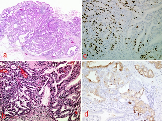
A 60-year-old man presented with melena and hematemesis in 1984. Esophagogastroduodenoscopy (EGD) detected a small protruding lesion in the duodenal bulb, which was diagnosed as Brunner's adenoma. No significant change was detected in subsequent annual EGD and biopsies for 10 years, after which the patient was not observed for 7 years. The patient presented with melena again in 2001. The lesion had changed shape to become a 10 mm sessile tumor with a central depression, and following a biopsy was diagnosed as an adenocarcinoma. The patient underwent partial resection of the duodenum. Histopathological assessment showed acidophilic cells with swollen nuclei, and clear cells forming a tubular or papillary tubule in the mucosal lamina propria and submucosal layer. The tumor cells stained positive for lysozyme, indicating that they arose from Brunner's gland. The patient showed no sign of recurrence and was disease-free for more than 34 months after surgery. The patient died of pneumonia. This is an extremely rare case of primary duodenal carcinoma arising from Brunner's gland in a patient observed for 17 years.
一名60岁男性于1984年出现黑便和呕血症状。食管胃十二指肠镜检查(EGD)发现十二指肠球部有一个小的突出病变,诊断为布伦纳腺瘤。在随后的10年中,每年进行的EGD和活检均未发现明显变化,此后7年未对该患者进行观察。2001年,该患者再次出现黑便。病变形状已变为一个10毫米的无蒂肿瘤,中央有凹陷,活检后诊断为腺癌。患者接受了十二指肠部分切除术。组织病理学评估显示,嗜酸性细胞细胞核肿胀,在黏膜固有层和黏膜下层可见透明细胞形成管状或乳头状小管。肿瘤细胞溶菌酶染色呈阳性,表明它们起源于布伦纳腺。患者术后未出现复发迹象,术后34个月以上无疾病。患者死于肺炎。这是一例极为罕见的起源于布伦纳腺的原发性十二指肠癌病例,对该患者进行了17年的观察。