Neuroscience Training Program, University of Wisconsin-Madison, Madison, WI 53705, USA.
Neuroimage. 2011 Jun 15;56(4):2129-37. doi: 10.1016/j.neuroimage.2011.03.074. Epub 2011 Apr 8.
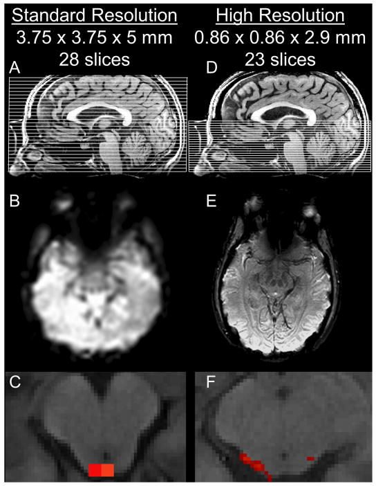
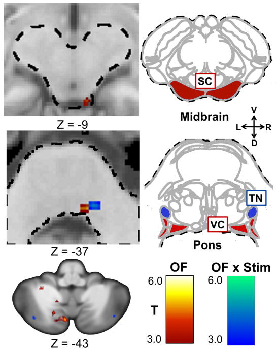
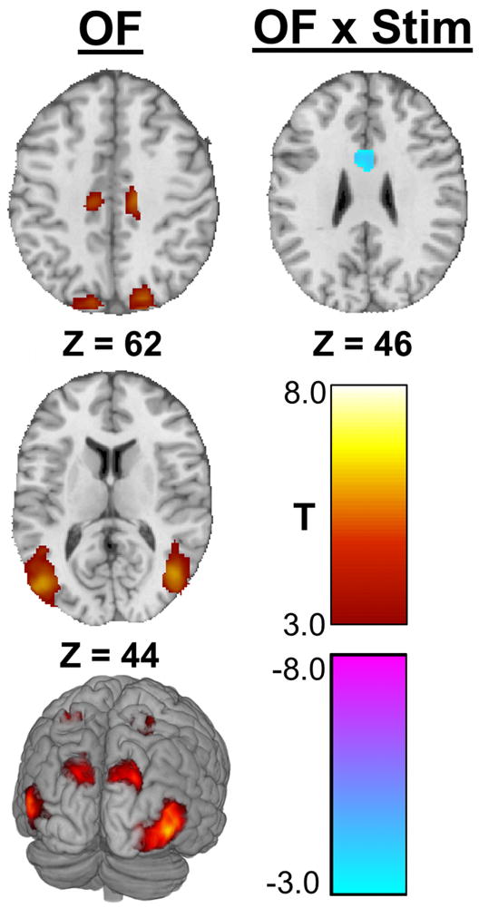
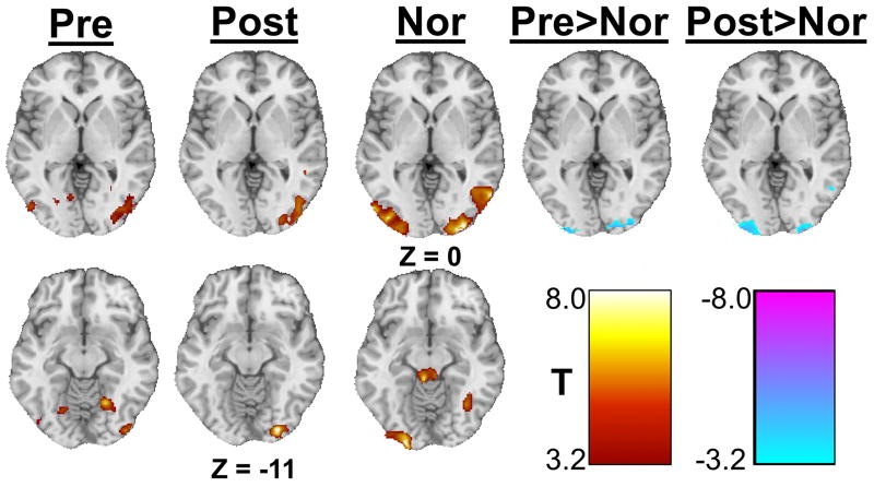
High-resolution functional magnetic resonance imaging (fMRI) can be used to precisely identify blood oxygen level dependent (BOLD) activation of small structures within the brainstem not accessible with standard fMRI. A previous study identified a region within the pons exhibiting sustained neuromodulation due to electrical tongue stimulation, but was unable to precisely identify the neuronal structure involved. For this study, high-resolution images of neural activity induced by optic flow were acquired in nine healthy controls and nine individuals with balance dysfunction before and after information-free tongue stimulation. Subjects viewed optic flow videos to activate the structures of interest. Sub-millimeter in-plane voxels of structures within the posterior fossa were acquired using a restricted field of view. Whole-brain functional imaging verified that global activation patterns due to optic flow were consistent with previous studies. Optic flow activated the visual association cortices, the vestibular nuclei, and the superior colliculus, as well as multiple regions within the cerebellum. The anterior cingulate cortex showed decreased activity after stimulation, while a region within the pons had increased post-stimulation activity. These observations suggest the pontine region is the trigeminal nucleus and that tongue stimulation interfaces with the balance-processing network within the pons. This high-resolution imaging allows detection of activity within individual brainstem nuclei not possible using standard resolution imaging.
高分辨率功能磁共振成像(fMRI)可用于精确识别脑干内标准 fMRI 无法触及的小结构的血氧水平依赖(BOLD)激活。先前的一项研究确定了脑桥内由于电舌刺激而持续表现出神经调节的区域,但无法精确确定涉及的神经元结构。在这项研究中,在信息自由舌刺激之前和之后,在 9 名健康对照者和 9 名平衡功能障碍者中获得了由光流引起的神经活动的高分辨率图像。受检者观看光流视频以激活感兴趣的结构。使用受限视野获取后颅窝内结构的亚毫米平面体素。全脑功能成像证实,由于光流引起的全局激活模式与先前的研究一致。光流激活了视觉联合皮质、前庭核和上丘,以及小脑内的多个区域。刺激后前扣带皮层的活动减少,而脑桥内的一个区域刺激后的活动增加。这些观察结果表明,脑桥区域是三叉神经核,并且舌刺激与脑桥内的平衡处理网络相互作用。这种高分辨率成像允许检测使用标准分辨率成像不可能检测到的单个脑干核内的活动。