• 文献检索
  • 文档翻译
  • 深度研究
  • 学术资讯
  • Suppr Zotero 插件Zotero 插件
  • 邀请有礼
  • 套餐&价格
  • 历史记录
应用&插件
Suppr Zotero 插件Zotero 插件浏览器插件Mac 客户端Windows 客户端微信小程序
定价
高级版会员购买积分包购买API积分包
服务
文献检索文档翻译深度研究API 文档MCP 服务
关于我们
关于 Suppr公司介绍联系我们用户协议隐私条款
关注我们

Suppr 超能文献

核心技术专利:CN118964589B侵权必究
粤ICP备2023148730 号-1Suppr @ 2026

文献检索

告别复杂PubMed语法,用中文像聊天一样搜索,搜遍4000万医学文献。AI智能推荐,让科研检索更轻松。

立即免费搜索

文件翻译

保留排版,准确专业,支持PDF/Word/PPT等文件格式,支持 12+语言互译。

免费翻译文档

深度研究

AI帮你快速写综述,25分钟生成高质量综述,智能提取关键信息,辅助科研写作。

立即免费体验

口腔扁平苔藓与口腔苔藓样病变中IgG细胞数量及分布的比较研究

Comparative study of number and distribution of IgG cells in oral lichen planus and oral lichenoid lesions.

作者信息

Ghalayani Parichehr, Razavi Seyed Mohammed, Gholami Darab

机构信息

Associate Professor, Department of Oral Medicine and Torabinejad Dental Research Center, School of Dentistry, Isfahan University of Medical Sciences, Isfahan, Iran.

出版信息

Dent Res J (Isfahan). 2009 Spring;6(1):1-5.

PMID:21528022
原文链接:https://pmc.ncbi.nlm.nih.gov/articles/PMC3075445/
Abstract

BACKGROUND

Oral lichen planus is a common mucocutaneous disorder with unknown etiology. While current data suggest that oral lichen planus is a cell-mediated disease, differential diagnosis of this disease and oral lichenoid lesions is very problematic, both clinically and histopathologically. This study aimed to compare immunohistochemical features of these similar diseases.

METHODS

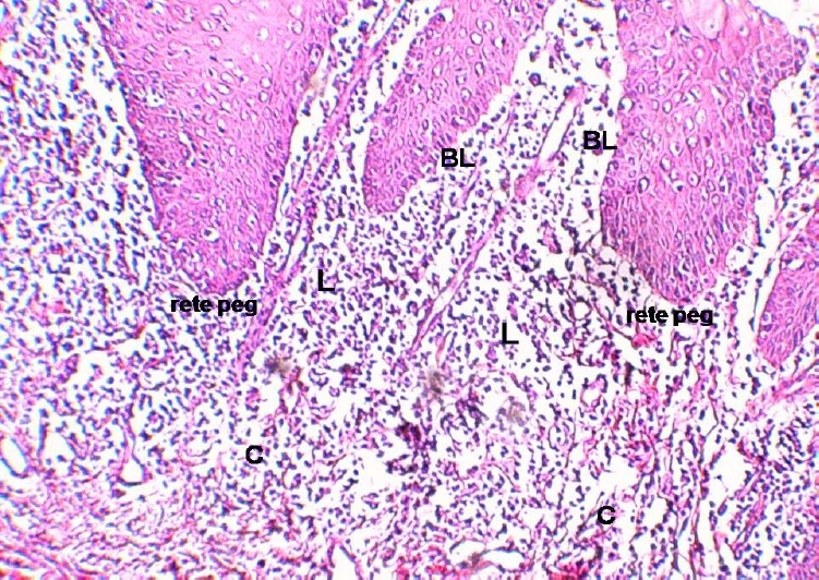
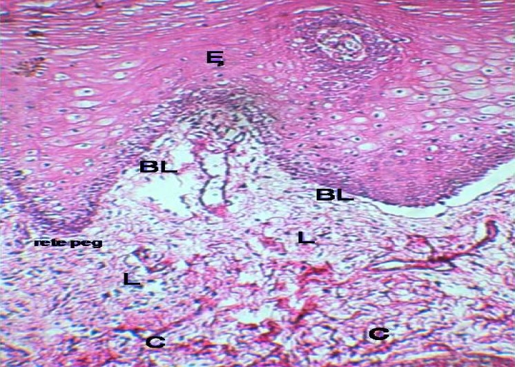
This was a descriptive-analytic study in which formalin-fixed and paraffin-embedded tissue sections of 30 oral lichen planus and 30 oral lichenoid lesions were immunohistochemically analyzed for number and distribution of IgG(+) cells. A standard biotin-streptavidin procedure after antigen retrieval was used. Data were analyzed in SPSS software using Mann-Whitney U test.

RESULTS

There were some significant differences in distribution of IgG (+)cells among different locations in oral lichen planus and also in oral lichenoid lesions separately; but the differences between distribution of IgG(+) cells between the two groups of oral lichen planus and oral lichenoid lesions were not significant.

CONCLUSION

There was no significant difference in number and distribution of IgG(+) cells between the two groups. So, this study can suggest that location of IgG is similar in samples of oral lichen planus and oral lichenoid lesions and consequently, this marker cannot help us differentiate them from each other. Other markers can be analyzed in further studies in order to find an appropriate distinguisher between the two lesions.

摘要

背景

口腔扁平苔藓是一种病因不明的常见黏膜皮肤疾病。虽然目前的数据表明口腔扁平苔藓是一种细胞介导的疾病,但在临床和组织病理学上,该疾病与口腔苔藓样病变的鉴别诊断都非常困难。本研究旨在比较这些相似疾病的免疫组化特征。

方法

这是一项描述性分析研究,对30例口腔扁平苔藓和30例口腔苔藓样病变的福尔马林固定石蜡包埋组织切片进行免疫组化分析,以检测IgG(+)细胞的数量和分布。采用抗原修复后的标准生物素-链霉亲和素程序。使用SPSS软件通过曼-惠特尼U检验对数据进行分析。

结果

口腔扁平苔藓不同部位以及口腔苔藓样病变不同部位的IgG(+)细胞分布存在一些显著差异;但口腔扁平苔藓组和口腔苔藓样病变组之间IgG(+)细胞分布的差异不显著。

结论

两组之间IgG(+)细胞的数量和分布没有显著差异。因此,本研究表明口腔扁平苔藓和口腔苔藓样病变样本中IgG的定位相似,因此,该标志物无法帮助我们区分它们。在进一步的研究中可以分析其他标志物,以便找到区分这两种病变的合适方法。

https://cdn.ncbi.nlm.nih.gov/pmc/blobs/e781/3075445/f44f8bdcdb1a/DRJ-6-1-g002.jpg
https://cdn.ncbi.nlm.nih.gov/pmc/blobs/e781/3075445/64b00eff347d/DRJ-6-1-g001.jpg
https://cdn.ncbi.nlm.nih.gov/pmc/blobs/e781/3075445/f44f8bdcdb1a/DRJ-6-1-g002.jpg
https://cdn.ncbi.nlm.nih.gov/pmc/blobs/e781/3075445/64b00eff347d/DRJ-6-1-g001.jpg
https://cdn.ncbi.nlm.nih.gov/pmc/blobs/e781/3075445/f44f8bdcdb1a/DRJ-6-1-g002.jpg

相似文献

1
Comparative study of number and distribution of IgG cells in oral lichen planus and oral lichenoid lesions.口腔扁平苔藓与口腔苔藓样病变中IgG细胞数量及分布的比较研究
Dent Res J (Isfahan). 2009 Spring;6(1):1-5.
2
Histomorphometric analysis of nuclear and cellular volumetric alterations in oral lichen planus, lichenoid lesions and normal oral mucosa using image analysis software.使用图像分析软件对口腔扁平苔藓、苔藓样病变和正常口腔黏膜中的细胞核及细胞体积变化进行组织形态计量分析。
Indian J Dent Res. 2013 Mar-Apr;24(2):277. doi: 10.4103/0970-9290.116678.
3
CD44 Expression in Oral Lichen Planus and Related Lesions-An Immunohistochemical Study.口腔扁平苔藓及相关病变中CD44的表达——一项免疫组织化学研究
Indian Dermatol Online J. 2023 Aug 29;14(5):624-629. doi: 10.4103/idoj.idoj_702_22. eCollection 2023 Sep-Oct.
4
Evaluation of mast cells, eosinophils, blood capillaries in oral lichen planus and oral lichenoid mucositis.口腔扁平苔藓和口腔苔藓样黏膜炎中肥大细胞、嗜酸性粒细胞及毛细血管的评估
Indian J Dent Res. 2012 Sep-Oct;23(5):695-6. doi: 10.4103/0970-9290.107422.
5
Histopathologic evaluation of oral lichen planus and oral lichenoid reaction: A comparative analysis based on basement membrane thickness and the distribution of mast cells.口腔扁平苔藓和口腔苔藓样反应的组织病理学评估:基于基底膜厚度和肥大细胞分布的比较分析
J Oral Maxillofac Pathol. 2021 Sep-Dec;25(3):549-550. doi: 10.4103/jomfp.JOMFP_220_20. Epub 2022 Jan 11.
6
Senescence in oral lichen planus as assessed by the immunohistochemical evaluation of senescence marker protein-30 (Regucalcin).通过免疫组织化学评估衰老标志物蛋白-30(Regucalcin)评估口腔扁平苔藓中的衰老。
Indian J Pathol Microbiol. 2023 Jan-Mar;66(1):9-13. doi: 10.4103/ijpm.ijpm_864_21.
7
Role of Mast Cells in Oral Lichen Planus and Oral Lichenoid Reactions.肥大细胞在口腔扁平苔藓和口腔苔藓样反应中的作用。
Autoimmune Dis. 2018 Jan 17;2018:7936564. doi: 10.1155/2018/7936564. eCollection 2018.
8
Histopathology of oral lichen planus and oral lichenoid lesions: An exploratory cross-sectional study.口腔扁平苔藓和口腔苔藓样病变的组织病理学:一项探索性横断面研究。
Oral Dis. 2023 Apr;29(3):1259-1268. doi: 10.1111/odi.14112. Epub 2022 Jan 4.
9
Oral lichen planus: A disease or a spectrum of tissue reactions? Types, causes, diagnostic algorhythms, prognosis, management strategies.口腔扁平苔藓:一种疾病还是一系列组织反应?类型、病因、诊断算法、预后、治疗策略。
Periodontol 2000. 2019 Jun;80(1):105-125. doi: 10.1111/prd.12260.
10
Histochemical analysis of pathological alterations in oral lichen planus and oral lichenoid lesions.口腔扁平苔藓和口腔苔藓样病变病理改变的组织化学分析
J Oral Sci. 2006 Dec;48(4):185-93. doi: 10.2334/josnusd.48.185.

引用本文的文献

1
Oral Lichen Planus: an Overview of Potential Risk Factors, Biomarkers and Treatments.口腔扁平苔藓:潜在风险因素、生物标志物及治疗概述
Asian Pac J Cancer Prev. 2018 May 26;19(5):1161-1167. doi: 10.22034/APJCP.2018.19.5.1161.
2
Oral lichenoid lesions - a review and update.口腔苔藓样病变——综述与更新
Indian J Dermatol. 2015 Jan-Feb;60(1):102. doi: 10.4103/0019-5154.147830.
3
Salivary IgA and IgG in oral lichen planus and oral lichenoid reactions diseases.口腔扁平苔藓和口腔苔藓样反应性疾病中的唾液免疫球蛋白A和免疫球蛋白G

本文引用的文献

1
Serum immunoglobulins IgG, IgA and IgM in patients with oral lichen ruber.口腔扁平苔藓患者的血清免疫球蛋白IgG、IgA和IgM
Coll Antropol. 2008 Mar;32(1):161-3.
2
Oral lichenoid lesions and allergy to dental materials.口腔苔藓样病变与牙科材料过敏
Biomed Pap Med Fac Univ Palacky Olomouc Czech Repub. 2007 Dec;151(2):333-9. doi: 10.5507/bp.2007.057.
3
Amalgam-tattoo-associated oral lichenoid lesion.
Contact Dermatitis. 2007 Apr;56(4):240-1. doi: 10.1111/j.1600-0536.2007.00915.x.
Adv Biomed Res. 2012;1:73. doi: 10.4103/2277-9175.102977. Epub 2012 Oct 31.
4
Cys-X-Cys ligand 9 might be an immunological factor in the pathogenesis of oral submucous fibrosis and its concomitant oral lichenoid lesion.Cys-X-Cys 配体 9 可能是口腔黏膜下纤维性变及其伴发性口腔扁平苔藓发病机制中的一个免疫学因素。
Clin Oral Investig. 2013 May;17(4):1251-8. doi: 10.1007/s00784-012-0799-9. Epub 2012 Jul 21.
4
The possible premalignant character of oral lichen planus and oral lichenoid lesions: a prospective five-year follow-up study of 192 patients.口腔扁平苔藓和口腔苔藓样病变的潜在癌前特征:192例患者的前瞻性五年随访研究
Oral Oncol. 2007 Sep;43(8):742-8. doi: 10.1016/j.oraloncology.2006.09.006. Epub 2006 Nov 16.
5
Lack of clinicopathologic correlation in the diagnosis of oral lichen planus based on the presently available diagnostic criteria and suggestions for modifications.基于目前可用诊断标准的口腔扁平苔藓诊断中临床病理相关性的缺乏及修改建议
J Oral Pathol Med. 2003 Oct;32(9):507-12. doi: 10.1034/j.1600-0714.2003.00125.x.
6
Oral lichen planus: causes, diagnosis and management.口腔扁平苔藓:病因、诊断与治疗
Aust Dent J. 2002 Dec;47(4):290-7. doi: 10.1111/j.1834-7819.2002.tb00540.x.
7
Immunohistochemical study of oral lichen planus associated with hepatitis C virus infection, oral lichenoid contact sensitivity reaction and idiopathic oral lichen planus.丙型肝炎病毒感染相关的口腔扁平苔藓、口腔苔藓样接触性敏感反应及特发性口腔扁平苔藓的免疫组织化学研究
Oral Dis. 2001 Sep;7(5):296-305. doi: 10.1034/j.1601-0825.2001.00718.x.
8
Immunofluorescence in oral lichen planus and oral lichenoid reaction. A review.口腔扁平苔藓和口腔苔藓样反应中的免疫荧光。综述。
Indian J Dent Res. 2001 Jan-Mar;12(1):29-34.
9
Lichen planus pemphigoides with circulating autoantibodies against 200 and 180 kDa epidermal antigens.伴有针对200kDa和180kDa表皮抗原的循环自身抗体的类天疱疮样扁平苔藓。
Eur J Dermatol. 2000 Apr-May;10(3):212-4.
10
[Lichen pemphigoid associated with developing hepatitis B in a child].[儿童中与乙型肝炎发生相关的类天疱疮样扁平苔藓]
Ann Dermatol Venereol. 1999 Aug-Sep;126(8-9):604-7.