Allendoerfer K L, Shelton D L, Shooter E M, Shatz C J
Department of Neurobiology, Stanford University School of Medicine, CA 94305.
Proc Natl Acad Sci U S A. 1990 Jan;87(1):187-90. doi: 10.1073/pnas.87.1.187.
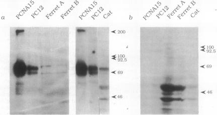
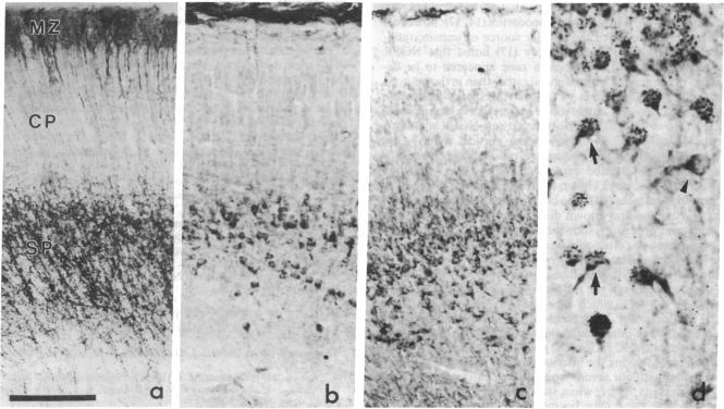
Nerve growth factor and its receptor (NGFR) are known to be present in diverse embryonic and neonatal central nervous system tissues, including the cerebral cortex. However, the identity of the cortical cells expressing NGFR immunoreactivity has not been established. We have used immunolabeling coupled with [3H]thymidine autoradiography to identify such cells in ferret and cat brain. Polyclonal antibodies raised against a synthetic peptide corresponding to a conserved amino acid sequence of the NGFR were used for this purpose. Western (immunologic) blot analyses show that these antibodies specifically recognize NGFR and precursor proteins. In both species, NGFR immunoreactivity is primarily associated with the early generated and transient subplate neuron population of the developing neocortex, as indicated by the following evidence: the immunoreactive cells (i) are located directly beneath the developing cortical plate, (ii) frequently have the inverted pyramid shape characteristic of subplate neurons, and (iii) can be labeled by an injection of [3H]thymidine on embryonic day (E) 28, a time when only subplate neurons are being generated. Intense NGFR immunostaining is seen on the cell bodies of these neurons as early as E30, several days after their last round of cell division, and this immunostaining remains strong for approximately 3 weeks. The NGFR immunoreactivity begins to decline around E52 and has disappeared from the region altogether by E60, at which time subplate neurons begin to die. The cellular localization and timing of expression suggest that the NGFR may play a role in the maintenance of subplate neurons and in the maturation of the cerebral cortex.
已知神经生长因子及其受体(NGFR)存在于多种胚胎和新生中枢神经系统组织中,包括大脑皮层。然而,表达NGFR免疫反应性的皮层细胞的身份尚未确定。我们使用免疫标记结合[3H]胸腺嘧啶放射自显影术来鉴定雪貂和猫脑中的此类细胞。为此,使用了针对与NGFR保守氨基酸序列相对应的合成肽产生的多克隆抗体。蛋白质免疫印迹分析表明,这些抗体能特异性识别NGFR和前体蛋白。在这两个物种中,NGFR免疫反应性主要与发育中的新皮层早期产生的、短暂的板下层神经元群体相关,如下证据所示:免疫反应性细胞(i)位于发育中的皮质板下方,(ii)通常具有板下层神经元特有的倒金字塔形状,(iii)在胚胎第28天注射[3H]胸腺嘧啶时可被标记,此时只有板下层神经元正在产生。早在胚胎第30天,即这些神经元最后一轮细胞分裂后的几天,就可以在其细胞体上看到强烈的NGFR免疫染色,这种免疫染色在大约3周内一直很强。NGFR免疫反应性在胚胎第52天左右开始下降,并在胚胎第60天从该区域完全消失,此时板下层神经元开始死亡。细胞定位和表达时间表明,NGFR可能在板下层神经元的维持和大脑皮层的成熟中发挥作用。