• 文献检索
  • 文档翻译
  • 深度研究
  • 学术资讯
  • Suppr Zotero 插件Zotero 插件
  • 邀请有礼
  • 套餐&价格
  • 历史记录
应用&插件
Suppr Zotero 插件Zotero 插件浏览器插件Mac 客户端Windows 客户端微信小程序
定价
高级版会员购买积分包购买API积分包
服务
文献检索文档翻译深度研究API 文档MCP 服务
关于我们
关于 Suppr公司介绍联系我们用户协议隐私条款
关注我们

Suppr 超能文献

核心技术专利:CN118964589B侵权必究
粤ICP备2023148730 号-1Suppr @ 2026

文献检索

告别复杂PubMed语法,用中文像聊天一样搜索,搜遍4000万医学文献。AI智能推荐,让科研检索更轻松。

立即免费搜索

文件翻译

保留排版,准确专业,支持PDF/Word/PPT等文件格式,支持 12+语言互译。

免费翻译文档

深度研究

AI帮你快速写综述,25分钟生成高质量综述,智能提取关键信息,辅助科研写作。

立即免费体验

鉴别腮腺良恶性肿瘤:5 分钟延迟的低剂量多期 CT 方案。

Distinguishing benign from malignant parotid gland tumours: low-dose multi-phasic CT protocol with 5-minute delay.

机构信息

Department of Radiology, Oncology Hospital, Guangxi Medical University, Nanning, People's Republic of China.

出版信息

Eur Radiol. 2011 Aug;21(8):1692-8. doi: 10.1007/s00330-011-2101-y. Epub 2011 Mar 11.

DOI:10.1007/s00330-011-2101-y
PMID:21547526
原文链接:https://pmc.ncbi.nlm.nih.gov/articles/PMC3128264/
Abstract

OBJECTIVES

To explore the percentage enhancement wash-out ratio (PEW) and relative PEW (RPEW) of low-dose multi-phasic computed tomography (CT) in distinguishing benign from malignant parotid gland tumours.

METHODS

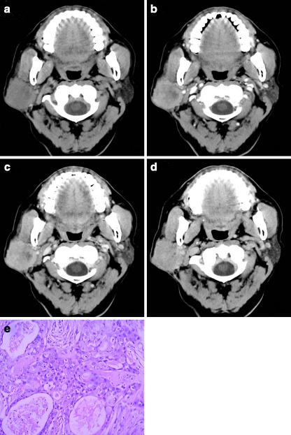
This study was approved by the ethics committee, and informed patient consent was obtained. 51 patients with parotid tumours proven by histopathology received CT, including 18 with pleomorphic adenomas, 14 with Warthin's tumours and 19 with malignant tumours. Size and attenuation of parotid tumours were measured. Compared with 5-min attenuation, the 30-s and 90-s PEW (PEW(30,) PEW(90)) and RPEW (RPEW(30), RPEW(90)) were calculated.

RESULTS

There was a significant difference in PEW(30), RPEW(30), PEW(90) and RPEW(90) in the parotid neoplasms groups (P < 0.01), and statistical significance existed simultaneously in pleomorphic adenomas vs malignant tumours and Warthin's tumours vs malignant tumours according to SNK-q test. The optimal diagnosis results of malignancy with 100% specificity (32/32) was obtained by using a combination of the following criteria: -70% > PEW(30) < 36%, -30% > PEW(30) < 19%, PEW(90) > 12%, and the sensitivity (74%) for diagnosis of malignancy was yield.

CONCLUSIONS

Wash-out ratio may assist in differentiating the benign from malignant parotid gland tumours. Combining the percentage of enhanced wash-out ratios of CT protocols can yield diagnostic results for malignancy.

摘要

目的

探讨低剂量多期 CT (CT)在鉴别腮腺良恶性肿瘤中的百分比增强洗脱率(PEW)和相对 PEW(RPEW)。

方法

本研究经伦理委员会批准,并获得患者知情同意。51 例经组织病理学证实的腮腺肿瘤患者接受 CT 检查,其中多形性腺瘤 18 例,Warthin 瘤 14 例,恶性肿瘤 19 例。测量腮腺肿瘤的大小和衰减。与 5 分钟衰减相比,计算 30 秒和 90 秒的 PEW(PEW(30),PEW(90))和 RPEW(RPEW(30),RPEW(90))。

结果

腮腺肿瘤组的 PEW(30)、RPEW(30)、PEW(90)和 RPEW(90)差异有统计学意义(P<0.01),SNK-q 检验显示多形性腺瘤与恶性肿瘤、Warthin 瘤与恶性肿瘤之间同时存在统计学意义。使用以下标准的组合,可获得 100%特异性(32/32)的恶性肿瘤最佳诊断结果:-70%>PEW(30)>-36%,-30%>PEW(30)>-19%,PEW(90)>12%,且诊断恶性肿瘤的敏感性(74%)。

结论

洗脱率可辅助鉴别腮腺良恶性肿瘤。结合 CT 方案的增强洗脱率百分比可以得出恶性肿瘤的诊断结果。

https://cdn.ncbi.nlm.nih.gov/pmc/blobs/ba24/3128264/e1c108d646d3/330_2011_2101_Fig1_HTML.jpg

相似文献

1
Distinguishing benign from malignant parotid gland tumours: low-dose multi-phasic CT protocol with 5-minute delay.鉴别腮腺良恶性肿瘤:5 分钟延迟的低剂量多期 CT 方案。
Eur Radiol. 2011 Aug;21(8):1692-8. doi: 10.1007/s00330-011-2101-y. Epub 2011 Mar 11.
2
Diagnostic value of CT perfusion imaging for parotid neoplasms.CT 灌注成像对腮腺肿瘤的诊断价值。
Dentomaxillofac Radiol. 2014;43(1):20130237. doi: 10.1259/dmfr.20130237. Epub 2013 Nov 1.
3
Perfusion imaging of parotid gland tumours: usefulness of arterial spin labeling for differentiating Warthin's tumours.腮腺肿瘤的灌注成像:动脉自旋标记在鉴别沃辛瘤中的应用价值
Eur Radiol. 2015 Nov;25(11):3247-54. doi: 10.1007/s00330-015-3755-7. Epub 2015 Apr 29.
4
Parotid gland tumors: comparison of conventional and diffusion-weighted MRI findings with histopathological results.腮腺肿瘤:常规 MRI 与弥散加权 MRI 表现与组织病理学结果的对比研究。
Dentomaxillofac Radiol. 2021 May 1;50(4):20200391. doi: 10.1259/dmfr.20200391. Epub 2020 Dec 11.
5
Application of DTI and ARFI imaging in differential diagnosis of parotid tumours.扩散张量成像(DTI)和剪切波弹性成像(ARFI)成像在腮腺肿瘤鉴别诊断中的应用
Dentomaxillofac Radiol. 2016;45(6):20160100. doi: 10.1259/dmfr.20160100. Epub 2016 Jul 22.
6
Sonoelastography of parotid gland tumours: initial experience and identification of characteristic patterns.腮腺肿瘤的超声弹性成像:初步经验和特征性模式的识别。
Eur Radiol. 2012 May;22(5):947-56. doi: 10.1007/s00330-011-2344-7. Epub 2012 Jan 22.
7
Pleomorphic adenoma of parotid gland: delayed enhancement on computed tomography.腮腺多形性腺瘤:计算机断层扫描延迟强化
Dentomaxillofac Radiol. 2008 Dec;37(8):464-9. doi: 10.1259/dmfr/79964183.
8
Distinguishing adrenal adenomas from nonadenomas: combined use of diagnostic parameters of unenhanced and short 5-minute dynamic enhanced CT protocol.鉴别肾上腺腺瘤与非腺瘤:未增强及短时间5分钟动态增强CT方案诊断参数的联合应用
Radiology. 2009 Feb;250(2):474-81. doi: 10.1148/radiol.2502080302. Epub 2008 Nov 26.
9
Differentiation of pleomorphic adenoma and Warthin's tumor of the parotid gland: ultrasonographic features.腮腺多形性腺瘤与沃辛瘤的鉴别诊断:超声特征
Acta Radiol. 2014 Dec;55(10):1203-9. doi: 10.1177/0284185113515865. Epub 2013 Dec 9.
10
Parotid tumors: differentiation of benign and malignant tumors with quantitative sonographic analyses.腮腺肿瘤:通过定量超声分析鉴别良性和恶性肿瘤
Ultrasound Med Biol. 2004 May;30(5):567-74. doi: 10.1016/j.ultrasmedbio.2004.02.007.

引用本文的文献

1
Distinguishing atypical parotid carcinomas and pleomorphic adenomas based on multiphasic computed tomography radiomics nomogram: a multicenter study.基于多期计算机断层扫描影像组学列线图鉴别非典型腮腺癌和多形性腺瘤:一项多中心研究
Front Oncol. 2025 Aug 1;15:1625487. doi: 10.3389/fonc.2025.1625487. eCollection 2025.
2
Enhanced CT combined with texture analysis for differential diagnosis of pleomorphic adenoma and adenolymphoma.增强 CT 联合纹理分析在多形性腺瘤与腺淋巴瘤鉴别诊断中的应用。
BMC Med Imaging. 2023 Oct 27;23(1):169. doi: 10.1186/s12880-023-01129-9.
3
Comparison of Different Machine Models Based on Multi-Phase Computed Tomography Radiomic Analysis to Differentiate Parotid Basal Cell Adenoma From Pleomorphic Adenoma.

本文引用的文献

1
A case of lipomatous pleomorphic adenoma in the parotid gland: a case report.腮腺脂肪性多形性腺瘤 1 例报告
Diagn Pathol. 2009 Jun 4;4:16. doi: 10.1186/1746-1596-4-16.
2
Salivary gland tumors: a single institution experience in India.涎腺肿瘤:印度一家机构的经验
Br J Oral Maxillofac Surg. 2008 Dec;46(8):635-8. doi: 10.1016/j.bjoms.2008.03.020. Epub 2008 Jul 11.
3
The value of dynamic MRI studies in parotid tumors.动态磁共振成像研究在腮腺肿瘤中的价值。
基于多期计算机断层扫描影像组学分析的不同机器型号对腮腺基底细胞腺瘤与多形性腺瘤进行鉴别诊断的比较
Front Oncol. 2022 Jul 12;12:889833. doi: 10.3389/fonc.2022.889833. eCollection 2022.
4
Machine learning-based multiparametric traditional multislice computed tomography radiomics for improving the discrimination of parotid neoplasms.基于机器学习的多参数传统多层计算机断层扫描影像组学用于提高腮腺肿瘤的鉴别诊断能力
Mol Clin Oncol. 2021 Nov;15(5):245. doi: 10.3892/mco.2021.2407. Epub 2021 Sep 24.
5
Deep Learning for Differentiating Benign From Malignant Parotid Lesions on MR Images.基于磁共振成像的深度学习鉴别腮腺良恶性病变
Front Oncol. 2021 Jun 23;11:632104. doi: 10.3389/fonc.2021.632104. eCollection 2021.
6
A Preliminary Study of CT Texture Analysis for Characterizing Epithelial Tumors of the Parotid Gland.腮腺上皮性肿瘤CT纹理分析特征的初步研究
Cancer Manag Res. 2020 Apr 21;12:2665-2674. doi: 10.2147/CMAR.S245344. eCollection 2020.
7
Delayed enhancement in differential diagnosis of salivary gland neoplasm.延迟强化在涎腺肿瘤鉴别诊断中的应用
Gland Surg. 2019 Sep;8(Suppl 3):S130-S135. doi: 10.21037/gs.2019.03.03.
8
Basal cell adenoma and myoepithelioma of the parotid gland: patterns of enhancement at two-phase CT in comparison with Warthin tumor.腮腺基底细胞腺瘤和肌上皮瘤:与沃辛瘤相比,双期 CT 增强模式。
Diagn Interv Radiol. 2019 Jul;25(4):285-290. doi: 10.5152/dir.2019.18337.
9
Contrast-enhanced Multi-detector CT Examination of Parotid Gland Tumors: Determination of the Most Helpful Scanning Delay for Predicting Histologic Subtypes.腮腺肿瘤的对比增强多层螺旋CT检查:确定预测组织学亚型最有用的扫描延迟时间
J Belg Soc Radiol. 2019 Jan 3;103(1):2. doi: 10.5334/jbsr.1596.
10
Diagnostic value of single-source dual-energy spectral computed tomography in differentiating parotid gland tumors: initial results.单源双能量光谱计算机断层扫描在鉴别腮腺肿瘤中的诊断价值:初步结果
Quant Imaging Med Surg. 2018 Jul;8(6):588-596. doi: 10.21037/qims.2018.07.07.
Acad Radiol. 2007 Jun;14(6):701-10. doi: 10.1016/j.acra.2007.03.004.
4
Dynamic multislice computed tomography findings for parotid gland tumors.腮腺肿瘤的动态多层计算机断层扫描结果
J Comput Assist Tomogr. 2007 Mar-Apr;31(2):309-16. doi: 10.1097/01.rct.0000236418.82395.b3.
5
Dynamic CT Evaluation of Tumor Vascularity in Renal Cell Carcinoma.肾细胞癌肿瘤血管的动态CT评估
AJR Am J Roentgenol. 2006 May;186(5):1423-30. doi: 10.2214/AJR.04.1408.
6
Solitary pulmonary nodule: characterization with combined wash-in and washout features at dynamic multi-detector row CT.孤立性肺结节:在动态多排CT上通过结合注入期和洗脱期特征进行特征描述
Radiology. 2005 Nov;237(2):675-83. doi: 10.1148/radiol.2372041549.
7
Salivary gland tumors in a Brazilian population: a retrospective study of 496 cases.巴西人群中的唾液腺肿瘤:496例病例的回顾性研究。
Int J Oral Maxillofac Surg. 2005 Jul;34(5):533-6. doi: 10.1016/j.ijom.2005.02.005.
8
Warthin tumor: a common, benign tumor presenting as a highly suspicious mass.沃辛瘤:一种常见的良性肿瘤,表现为高度可疑的肿块。
J Am Board Fam Pract. 2005 Jul-Aug;18(4):320-2. doi: 10.3122/jabfm.18.4.320.
9
Warthin tumor of the parotid gland: diagnostic value of MR imaging with histopathologic correlation.腮腺沃辛瘤:磁共振成像与组织病理学相关性的诊断价值
AJNR Am J Neuroradiol. 2004 Aug;25(7):1256-62.
10
Parotid tumors: MR imaging with pathological correlation.腮腺肿瘤:磁共振成像与病理对照
Eur Radiol. 2003 Dec;13 Suppl 4:L25-33. doi: 10.1007/s00330-003-1999-0.