• 文献检索
  • 文档翻译
  • 深度研究
  • 学术资讯
  • Suppr Zotero 插件Zotero 插件
  • 邀请有礼
  • 套餐&价格
  • 历史记录
应用&插件
Suppr Zotero 插件Zotero 插件浏览器插件Mac 客户端Windows 客户端微信小程序
定价
高级版会员购买积分包购买API积分包
服务
文献检索文档翻译深度研究API 文档MCP 服务
关于我们
关于 Suppr公司介绍联系我们用户协议隐私条款
关注我们

Suppr 超能文献

核心技术专利:CN118964589B侵权必究
粤ICP备2023148730 号-1Suppr @ 2026

文献检索

告别复杂PubMed语法,用中文像聊天一样搜索,搜遍4000万医学文献。AI智能推荐,让科研检索更轻松。

立即免费搜索

文件翻译

保留排版,准确专业,支持PDF/Word/PPT等文件格式,支持 12+语言互译。

免费翻译文档

深度研究

AI帮你快速写综述,25分钟生成高质量综述,智能提取关键信息,辅助科研写作。

立即免费体验

腮腺上皮性肿瘤CT纹理分析特征的初步研究

A Preliminary Study of CT Texture Analysis for Characterizing Epithelial Tumors of the Parotid Gland.

作者信息

Zhang Dan, Li Xiaojiao, Lv Liang, Yu Jiayi, Yang Chao, Xiong Hua, Liao Ruikun, Zhou Bi, Huang Xianlong, Liu Xiaoshuang, Tang Zhuoyue

机构信息

Department of Radiology, Chongqing General Hospital, University of Chinese Academy of Sciences, Chongqing 400014, People's Republic of China.

Molecular and Functional Imaging Laboratory, Chongqing General Hospital, University of Chinese Academy of Sciences, Chongqing 400014, People's Republic of China.

出版信息

Cancer Manag Res. 2020 Apr 21;12:2665-2674. doi: 10.2147/CMAR.S245344. eCollection 2020.

DOI:10.2147/CMAR.S245344
PMID:32368145
原文链接:https://pmc.ncbi.nlm.nih.gov/articles/PMC7183330/
Abstract

OBJECTIVE

The aim of this study was to explore and validate the diagnostic performance of whole-volume CT texture features in differentiating the common benign and malignant epithelial tumors of the parotid gland.

MATERIALS AND METHODS

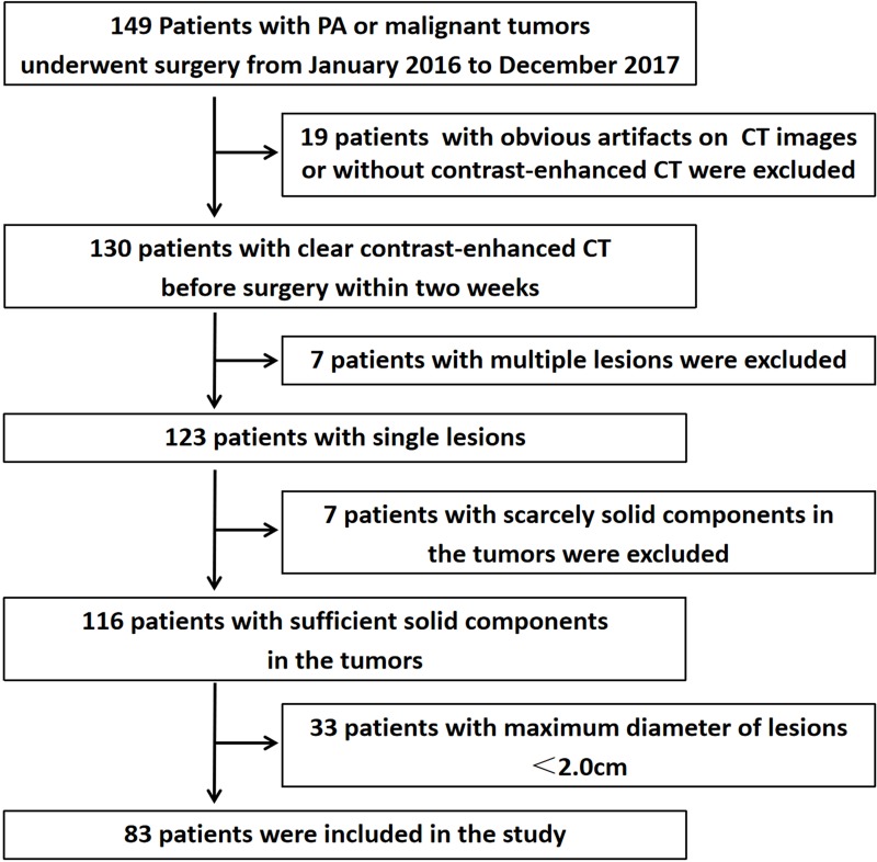
Contrast-enhanced CT images of 83 patients with common benign and malignant epithelial tumors of the parotid gland confirmed by histopathology were retrospectively analyzed, including 50 patients with pleomorphic adenoma (PA) and 33 patients with malignant epithelial tumors. Quantitative texture features of tumors were extracted from CT images of arterial phase. The diagnostic performance of texture features was evaluated via receiver operating characteristic (ROC) curve and area under ROC curve (AUC). The specificity and sensitivity were respectively discussed by the maximum Youden's index.

RESULTS

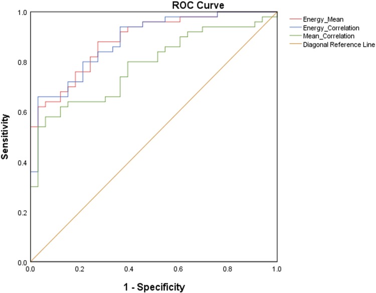
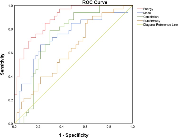
All the texture features were subject to normal distribution and homoscedasticity. Energy, mean, correlation, and sum entropy of epithelial malignancy group were significantly higher than those of PA group (P<0.05). There were no statistically significant differences between PA group and epithelial malignancy group in uniformity, entropy, skewness, kurtosis, contrast, and difference entropy (P>0.05). The AUC of each texture feature and joint diagnostic model was 0.887 (energy), 0.734 (mean), 0.739 (correlation), 0.623 (sum entropy), 0.888 (energy-mean), 0.883 (energy-correlation), 0.784 (mean-correlation). The diagnostic efficiency of energy-mean was the best. Based on the maximum Youden's index, the specificity of energy-correlation was the highest (97%) and the sensitivity of energy was the highest (97%).

CONCLUSION

Energy, mean, correlation, and sum entropy can be the effective quantitative texture features to differentiate the benign and malignant epithelial tumors of the parotid gland. With higher AUC, energy and energy-mean are superior to other indexes or joint diagnostic models in differentiating the benign and malignant epithelial tumors of the parotid gland. CT texture analysis can be used as a noninvasive and valuable means of preoperative assessment of parotid epithelial tumors without additional cost to the patients.

摘要

目的

本研究旨在探讨并验证全容积CT纹理特征在鉴别腮腺常见良性和恶性上皮性肿瘤中的诊断性能。

材料与方法

回顾性分析83例经组织病理学证实的腮腺常见良性和恶性上皮性肿瘤患者的增强CT图像,其中多形性腺瘤(PA)患者50例,恶性上皮性肿瘤患者33例。从动脉期CT图像中提取肿瘤的定量纹理特征。通过受试者操作特征(ROC)曲线和ROC曲线下面积(AUC)评估纹理特征的诊断性能。采用最大约登指数分别讨论特异性和敏感性。

结果

所有纹理特征均服从正态分布且具有方差齐性。上皮性恶性肿瘤组的能量、均值、相关性和总和熵均显著高于PA组(P<0.05)。PA组与上皮性恶性肿瘤组在均匀性、熵、偏度、峰度、对比度和差异熵方面无统计学差异(P>0.05)。各纹理特征及联合诊断模型的AUC分别为0.887(能量)、0.734(均值)、0.739(相关性)、0.623(总和熵)、0.888(能量-均值)、0.883(能量-相关性)、0.784(均值-相关性)。能量-均值的诊断效率最佳。基于最大约登指数,能量-相关性的特异性最高(97%),能量的敏感性最高(97%)。

结论

能量、均值、相关性和总和熵可作为鉴别腮腺良性和恶性上皮性肿瘤的有效定量纹理特征。能量和能量-均值在鉴别腮腺良性和恶性上皮性肿瘤方面具有较高的AUC,优于其他指标或联合诊断模型。CT纹理分析可作为一种无创且有价值的手段,用于腮腺上皮性肿瘤的术前评估,且无需患者额外付费。

https://cdn.ncbi.nlm.nih.gov/pmc/blobs/8070/7183330/3ee37c7b9a62/CMAR-12-2665-g0004.jpg
https://cdn.ncbi.nlm.nih.gov/pmc/blobs/8070/7183330/caf942beee7a/CMAR-12-2665-g0001.jpg
https://cdn.ncbi.nlm.nih.gov/pmc/blobs/8070/7183330/9556ecf97a16/CMAR-12-2665-g0002.jpg
https://cdn.ncbi.nlm.nih.gov/pmc/blobs/8070/7183330/bbccff24e5bd/CMAR-12-2665-g0003.jpg
https://cdn.ncbi.nlm.nih.gov/pmc/blobs/8070/7183330/3ee37c7b9a62/CMAR-12-2665-g0004.jpg

相似文献

1
A Preliminary Study of CT Texture Analysis for Characterizing Epithelial Tumors of the Parotid Gland.腮腺上皮性肿瘤CT纹理分析特征的初步研究
Cancer Manag Res. 2020 Apr 21;12:2665-2674. doi: 10.2147/CMAR.S245344. eCollection 2020.
2
Improving the diagnosis of common parotid tumors via the combination of CT image biomarkers and clinical parameters.通过 CT 图像生物标志物与临床参数的结合来改善常见腮腺肿瘤的诊断。
BMC Med Imaging. 2020 Apr 15;20(1):38. doi: 10.1186/s12880-020-00442-x.
3
Histogram analysis of ultrasonographic images in the differentiation of benign and malignant parotid gland tumors.超声图像直方图分析在腮腺良恶性肿瘤鉴别诊断中的价值。
Oral Surg Oral Med Oral Pathol Oral Radiol. 2023 Aug;136(2):240-246. doi: 10.1016/j.oooo.2023.04.011. Epub 2023 May 1.
4
Texture analysis in the characterization of parotid salivary gland lesions: A study on MR diffusion weighted imaging.腮腺唾液腺病变的纹理分析:MR 扩散加权成像研究。
Eur J Radiol. 2021 Mar;136:109529. doi: 10.1016/j.ejrad.2021.109529. Epub 2021 Jan 7.
5
Apparent diffusion coefficient (ADC) histogram analysis: differentiation of benign from malignant parotid gland tumors using readout-segmented diffusion-weighted imaging.表观扩散系数(ADC)直方图分析:应用分段读出扩散加权成像对腮腺良恶性肿瘤进行鉴别诊断。
Dentomaxillofac Radiol. 2019 Oct;48(7):20190100. doi: 10.1259/dmfr.20190100. Epub 2019 Jul 9.
6
Histogram analysis of dynamic contrast-enhanced magnetic resonance imaging in the differential diagnosis of parotid tumors.动态对比增强磁共振成像直方图分析在腮腺肿瘤鉴别诊断中的应用。
BMC Med Imaging. 2021 Dec 17;21(1):194. doi: 10.1186/s12880-021-00724-y.
7
[The value of high resolution diffusion weighted imaging in differentiating benign and malignant epithelial tumors of parotid gland].[高分辨率扩散加权成像在鉴别腮腺上皮性良恶性肿瘤中的价值]
Zhonghua Yi Xue Za Zhi. 2018 May 8;98(17):1347-1351. doi: 10.3760/cma.j.issn.0376-2491.2018.17.013.
8
Histogram analysis of apparent diffusion coefficient maps for differentiating malignant from benign parotid gland tumors.用于区分腮腺良恶性肿瘤的表观扩散系数图的直方图分析
Eur Arch Otorhinolaryngol. 2018 Aug;275(8):2151-2157. doi: 10.1007/s00405-018-5052-y. Epub 2018 Jul 2.
9
Texture-based and diffusion-weighted discrimination of parotid gland lesions on MR images at 3.0 Tesla.基于纹理和扩散加权的 3.0T MRI 腮腺病变鉴别。
NMR Biomed. 2013 Nov;26(11):1372-9. doi: 10.1002/nbm.2962. Epub 2013 May 23.
10
[Comparative analysis of computed tomography texture features between pulmonary inflammatory nodules and lung cancer].[肺炎性结节与肺癌的CT纹理特征对比分析]
Zhonghua Zhong Liu Za Zhi. 2018 Nov 23;40(11):847-850. doi: 10.3760/cma.j.issn.0253-3766.2018.11.010.

引用本文的文献

1
The relationship between radiomic features in CT images and the salivary gland SPECT/CT standardised uptake value using 3D analysis.使用三维分析的CT图像中放射组学特征与唾液腺SPECT/CT标准化摄取值之间的关系。
Pol J Radiol. 2025 May 23;90:e253-e259. doi: 10.5114/pjr/202590. eCollection 2025.
2
The value of multi-phase CT based intratumor and peritumoral radiomics models for evaluating capsular characteristics of parotid pleomorphic adenoma.基于多期CT的肿瘤内及瘤周影像组学模型在评估腮腺多形性腺瘤包膜特征中的价值
Front Med (Lausanne). 2025 Apr 22;12:1566555. doi: 10.3389/fmed.2025.1566555. eCollection 2025.
3

本文引用的文献

1
The prognostic value of CT-based image-biomarkers for head and neck cancer patients treated with definitive (chemo-)radiation.基于 CT 的影像学生物标志物对头颈部癌症患者接受根治性(放化疗)治疗的预后价值。
Oral Oncol. 2019 Aug;95:178-186. doi: 10.1016/j.oraloncology.2019.06.020. Epub 2019 Jun 26.
2
Tuberculosis of the parotid lymph nodes: clinical and imaging features.腮腺淋巴结结核:临床与影像学特征
Infect Drug Resist. 2018 Oct 11;11:1795-1805. doi: 10.2147/IDR.S164993. eCollection 2018.
3
Early Prediction of Acute Xerostomia During Radiation Therapy for Head and Neck Cancer Based on Texture Analysis of Daily CT.
Enhanced CT combined with texture analysis for differential diagnosis of pleomorphic adenoma and adenolymphoma.
增强 CT 联合纹理分析在多形性腺瘤与腺淋巴瘤鉴别诊断中的应用。
BMC Med Imaging. 2023 Oct 27;23(1):169. doi: 10.1186/s12880-023-01129-9.
4
Quantitative Analysis and Pathological Basis of Signal Intensity on T2-Weighted MR Images in Benign and Malignant Parotid Tumors.腮腺良恶性肿瘤T2加权磁共振图像信号强度的定量分析及病理基础
Cancer Manag Res. 2021 Jul 7;13:5423-5431. doi: 10.2147/CMAR.S319466. eCollection 2021.
5
The Role of Preoperative Computed Tomography Radiomics in Distinguishing Benign and Malignant Tumors of the Parotid Gland.术前计算机断层扫描影像组学在鉴别腮腺良恶性肿瘤中的作用
Front Oncol. 2021 Mar 10;11:634452. doi: 10.3389/fonc.2021.634452. eCollection 2021.
基于日常 CT 的纹理分析对头颈部癌症放射治疗中急性口干症的早期预测
Int J Radiat Oncol Biol Phys. 2018 Nov 15;102(4):1308-1318. doi: 10.1016/j.ijrobp.2018.04.059. Epub 2018 May 1.
4
Texture analysis as a predictor of radiation-induced xerostomia in head and neck patients undergoing IMRT.纹理分析作为预测头颈部调强放疗患者放射性口干的指标。
Radiol Med. 2018 Jun;123(6):415-423. doi: 10.1007/s11547-017-0850-7. Epub 2018 Jan 24.
5
Prediction of Pancreatic Neuroendocrine Tumor Grade Based on CT Features and Texture Analysis.基于 CT 特征和纹理分析预测胰腺神经内分泌肿瘤分级。
AJR Am J Roentgenol. 2018 Feb;210(2):341-346. doi: 10.2214/AJR.17.18417. Epub 2017 Nov 15.
6
Differential Diagnosis of Parotid Gland Tumors: Role of Shear Wave Elastography.腮腺肿瘤的鉴别诊断:剪切波弹性成像的作用。
Biomed Res Int. 2017;2017:9234672. doi: 10.1155/2017/9234672. Epub 2017 Sep 13.
7
Texture-based classification of different single liver lesion based on SPAIR T2W MRI images.基于SPAIR T2W MRI图像的不同单一肝脏病变的纹理分类
BMC Med Imaging. 2017 Jul 13;17(1):42. doi: 10.1186/s12880-017-0212-x.
8
Impact of Adjuvant Radiotherapy for Malignant Salivary Gland Tumors.辅助放疗对恶性涎腺肿瘤的影响
Otolaryngol Head Neck Surg. 2017 Dec;157(6):988-994. doi: 10.1177/0194599817717661. Epub 2017 Jul 4.
9
Surgical options in benign parotid tumors: a proposal for classification.腮腺良性肿瘤的手术选择:一种分类建议。
Eur Arch Otorhinolaryngol. 2017 Nov;274(11):3825-3836. doi: 10.1007/s00405-017-4650-4. Epub 2017 Jun 21.
10
Texture analysis of baseline multiphasic hepatic computed tomography images for the prognosis of single hepatocellular carcinoma after hepatectomy: A retrospective pilot study.肝切除术后单发性肝细胞癌预后的基线多期肝脏计算机断层扫描图像纹理分析:一项回顾性初步研究。
Eur J Radiol. 2017 May;90:198-204. doi: 10.1016/j.ejrad.2017.02.035. Epub 2017 Feb 23.