Department of Neurobiology, Yale University School of Medicine New Haven, CT, USA.
Front Neural Circuits. 2011 Apr 25;5:5. doi: 10.3389/fncir.2011.00005. eCollection 2011.
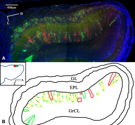
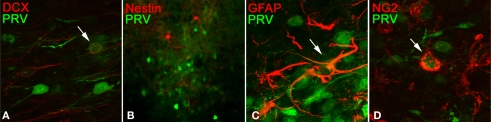
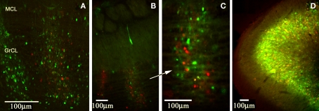
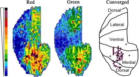
Lateral connections in the olfactory bulb were previously thought to be organized for center-surround inhibition. However, recent anatomical and physiological studies showed sparse and distributed interactions of inhibitory granule cells (GCs) which tended to be organized in columnar clusters. Little is known about how these distributed clusters are interconnected. In this study, we use transsynaptic tracing viruses bearing green or red fluorescent proteins to further elucidate mitral- and tufted-to-GC connectivity. Separate sites in the glomerular layer were injected with each virus. Columns with labeling from both viruses after transsynaptic spread show sparse red or green GCs which tended to be segregated. However, there was a higher incidence of co-labeled cells than chance would predict. Similar segregation of labeling is observed from dual injections into olfactory cortex. Collectively, these results suggest that neighboring mitral and tufted cells receive inhibitory inputs from segregated subsets of GCs, enabling inhibition of a center by specific and discontinuous lateral elements.
嗅球中的侧连接以前被认为是用于中心-环绕抑制的组织。然而,最近的解剖学和生理学研究显示,抑制性颗粒细胞(GCs)的稀疏和分布式相互作用往往呈柱状簇排列。关于这些分布式簇如何相互连接,人们知之甚少。在这项研究中,我们使用携带绿色或红色荧光蛋白的顺行示踪病毒,进一步阐明了僧帽细胞和丛状细胞与 GC 的连接。用每种病毒分别在肾小球层的不同部位注射。在顺行扩散后,来自两种病毒的标记的柱显示出稀疏的红色或绿色 GC,这些 GC 往往是分离的。然而,标记的细胞的出现频率高于随机预测。从双注射到嗅球皮层也观察到类似的标记分离。总的来说,这些结果表明,相邻的僧帽细胞和丛状细胞接收来自分离的 GC 子集的抑制性输入,从而使中心受到特定和不连续的侧部元件的抑制。