Institute for Antiviral Research, Department of Animal, Dairy, and Veterinary Sciences, Utah State University, Logan, Utah, United States of America.
PLoS One. 2011 May 9;6(5):e19575. doi: 10.1371/journal.pone.0019575.
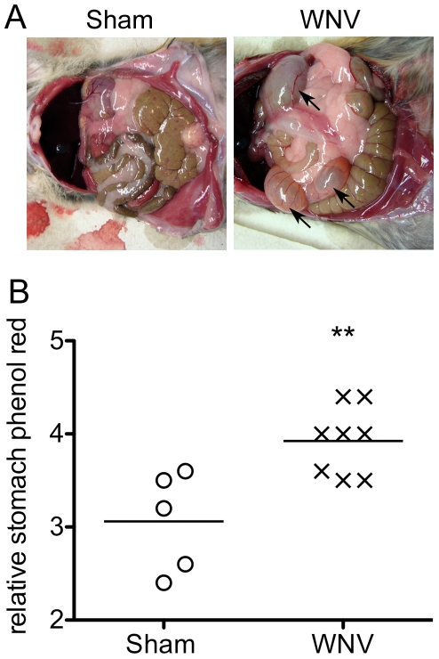
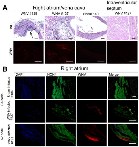
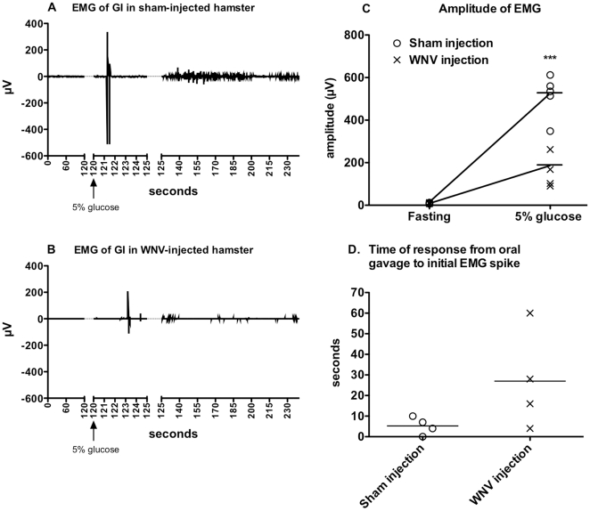
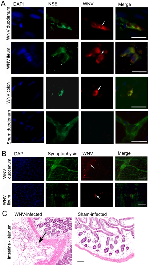
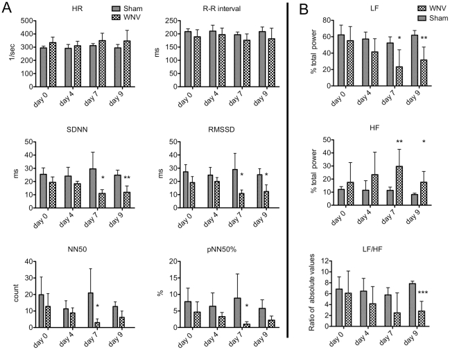
Clinical studies and case reports clearly document that West Nile virus (WNV) can cause respiratory and gastrointestinal (GI) complications. Other functions controlled by the autonomic nervous system may also be directly affected by WNV, such as bladder and cardiac functions. To investigate how WNV can cause autonomic dysfunctions, we focused on the cardiac and GI dysfunctions of rodents infected with WNV. Infected hamsters had distension of the stomach and intestines at day 9 after viral challenge. GI motility was detected by a dye retention assay; phenol red dye was retained more in the stomachs of infected hamsters as compared to sham-infected hamsters. The amplitudes of electromygraphs (EMGs) of intestinal muscles were significantly reduced. Myenteric neurons that innervate the intestines, in addition to neurons in the brain stem, were identified to be infected with WNV. These data suggest that infected neurons controlling autonomic function were the cause of GI dysfunction in WNV-infected hamsters. Using radiotelemetry to record electrocardiograms and to measure heart rate variability (HRV), a well-accepted readout for autonomic function, we determined that HRV and autonomic function were suppressed in WNV-infected hamsters. Cardiac histopathology was observed at day 9 only in the right atrium, which was coincident with WNV staining. A subset of WNV infected cells was identified among cells with hyperpolarization-activated cyclic nucleotide-gated potassium channel 4 (HCN4) as a marker for cells in the sinoatrial (SA) and atrioventricular (AV) nodes. The unique contribution of this study is the discovery that WNV infection of hamsters can lead to autonomic dysfunction as determined by reduced HRV and reduced EMG amplitudes of the GI tract. These data may model autonomic dysfunction of the human West Nile neurological disease.
临床研究和病例报告清楚地表明,西尼罗河病毒(WNV)可引起呼吸道和胃肠道(GI)并发症。自主神经系统控制的其他功能也可能直接受到 WNV 的影响,例如膀胱和心脏功能。为了研究 WNV 如何引起自主神经功能障碍,我们专注于感染 WNV 的啮齿动物的心脏和 GI 功能障碍。感染的仓鼠在病毒攻击后第 9 天胃和肠扩张。通过染料保留测定法检测 GI 蠕动;与假感染的仓鼠相比,感染的仓鼠胃中保留的苯酚红染料更多。肠肌电图(EMG)的幅度显着降低。除了脑干神经元外,支配肠道的神经元也被鉴定为感染了 WNV。这些数据表明,控制自主功能的感染神经元是 WNV 感染仓鼠 GI 功能障碍的原因。使用无线电遥测记录心电图并测量心率变异性(HRV),这是自主功能的一种公认的读数,我们确定 HRV 和自主功能在 WNV 感染的仓鼠中受到抑制。仅在第 9 天观察到右心房的心脏组织病理学,这与 WNV 染色一致。一组 HCN4 作为窦房结(SA)和房室结(AV)细胞标志物的超极化激活环核苷酸门控钾通道 4(HCN4)的细胞中鉴定出感染的 WNV 细胞。本研究的独特贡献是发现 WNV 感染仓鼠可导致自主神经功能障碍,其表现为 HRV 降低和 GI 道 EMG 幅度降低。这些数据可能模拟人类西尼罗河神经疾病的自主神经功能障碍。