Medical Research Council Human Reproductive Sciences Unit, The Queen's Medical Research Institute, University of Edinburgh, Edinburgh, United Kingdom.
PLoS One. 2011 May 12;6(5):e19209. doi: 10.1371/journal.pone.0019209.
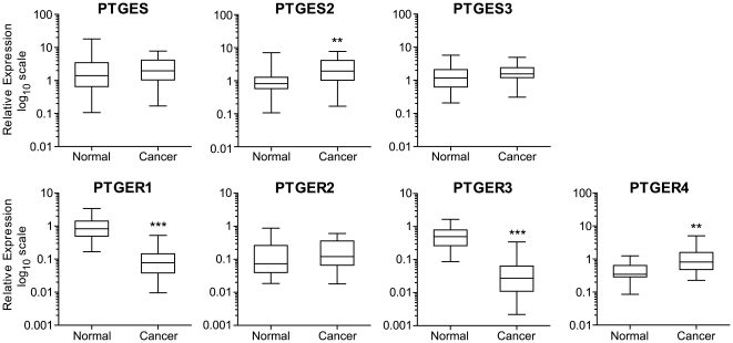
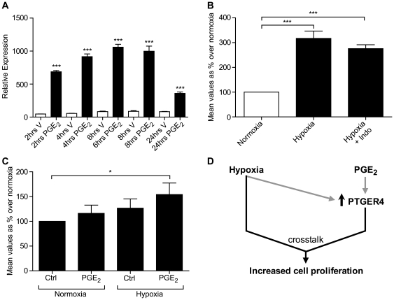
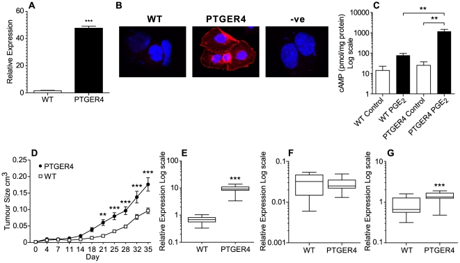
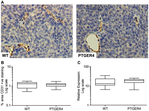
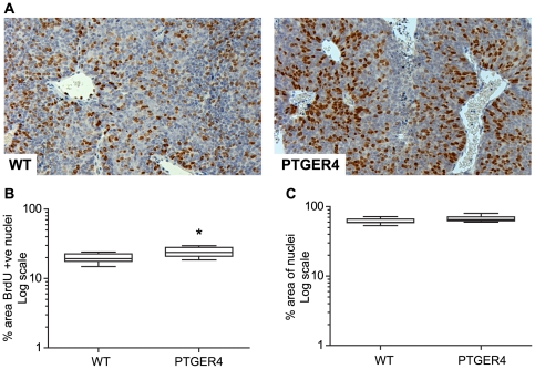
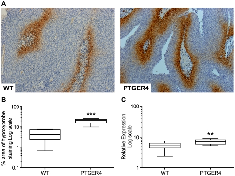
The prostaglandin endoperoxide synthase (PTGS) pathway is a potent driver of tumour development in humans by enhancing the biosynthesis and signalling of prostaglandin (PG) E(2). PTGS2 expression and PGE(2) biosynthesis is elevated in endometrial adenocarcinoma, however the mechanism whereby PTGS and PGE(2) regulate endometrial tumour growth is unknown. Here we investigated (a) the expression profile of the PGE synthase enzymes (PTGES, PTGES-2, PTGES-3) and PGE receptors (PTGER1-4) in endometrial adenocarcinomas compared with normal endometrium and (b) the role of PTGER4 in endometrial tumorigenesis in vivo. We found elevated expression of PTGES2 and PTGER4 and suppression of PTGER1 and PTGER3 in endometrial adenocarcinomas compared with normal endometrium. Using WT Ishikawa endometrial adenocarcinoma cells and Ishikawa cells stably transfected with the full length PTGER4 cDNA (PTGER4 cells) xenografted in the dorsal flanks of nude mice, we show that PTGER4 rapidly and significantly enhances tumour growth rate. Coincident with enhanced PTGER4-mediated tumour growth we found elevated expression of PTGS2 in PTGER4 xenografts compared with WT xenografts. Furthermore we found that the augmented growth rate of the PTGER4 xenografts was not due to enhanced angiogenesis, but regulated by an increased proliferation index and hypoxia. In vitro, we found that PGE(2) and hypoxia independently induce expression of PTGER4 indicating two independent pathways regulating prostanoid receptor expression. Finally we have shown that PGE(2) and hypoxia synergise to promote cellular proliferation of endometrial adenocarcinoma cells.
前列腺素内过氧化物合酶(PTGS)途径通过增强前列腺素(PG)E(2)的生物合成和信号转导,是人类肿瘤发展的有力驱动因素。PTGS2 表达和 PGE(2)生物合成在子宫内膜腺癌中升高,然而,PTGS 和 PGE(2)调节子宫内膜肿瘤生长的机制尚不清楚。在这里,我们研究了(a)与正常子宫内膜相比,PG 合酶酶(PTGES、PTGES-2、PTGES-3)和 PGE 受体(PTGER1-4)在子宫内膜腺癌中的表达谱;(b)PTGER4 在体内子宫内膜肿瘤发生中的作用。我们发现与正常子宫内膜相比,子宫内膜腺癌中 PTGES2 和 PTGER4 的表达升高,而 PTGER1 和 PTGER3 的表达受到抑制。我们使用 WT Ishikawa 子宫内膜腺癌细胞和稳定转染全长 PTGER4 cDNA 的 Ishikawa 细胞(PTGER4 细胞)异种移植到裸鼠背部侧翼,结果表明 PTGER4 可快速显著增强肿瘤生长速度。与增强的 PTGER4 介导的肿瘤生长一致,我们发现与 WT 异种移植物相比,PTGER4 异种移植物中 PTGS2 的表达升高。此外,我们发现 PTGER4 异种移植物的生长速度加快不是由于血管生成增强,而是由增殖指数和缺氧增加调节的。体外,我们发现 PGE(2)和缺氧独立诱导 PTGER4 的表达,表明有两种独立的途径调节前列腺素受体的表达。最后,我们已经表明 PGE(2)和缺氧协同促进子宫内膜腺癌细胞的增殖。