Department of Anesthesiology and Intensive Care Medicine, Tübingen University Hospital, Eberhard-Karls University Tübingen, Tübingen, Germany.
Inflammation. 2012 Apr;35(2):566-73. doi: 10.1007/s10753-011-9347-z.
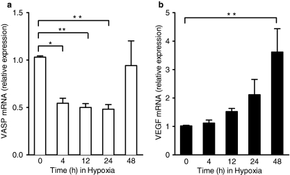
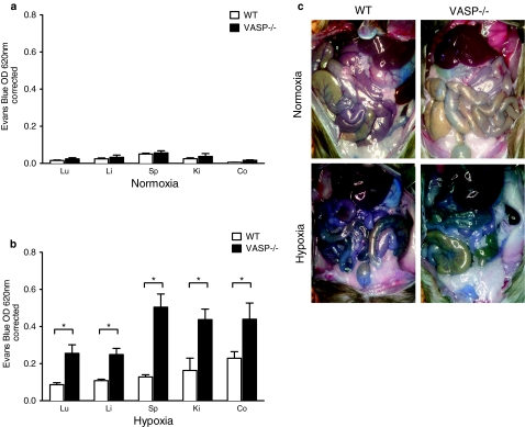
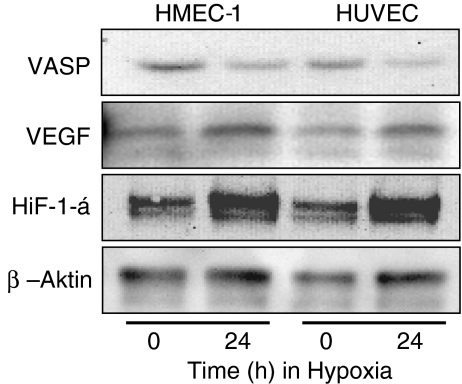
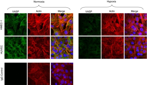
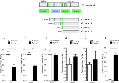
The endothelial barrier controls the passage of solutes from the vascular space. This is achieved through active reorganization of the actin cytoskeleton. A central cytoskeletal protein involved into this is vasodilator-stimulated phosphoprotein (VASP). However, the functional role of endothelial VASP during hypoxia has not been thoroughly elucidated. We determined endothelial VASP expression through real-time PCR (Rt-PCR), immunhistochemistry, and Western blot analysis during hypoxia. VASP promoter studies were performed using a PGL3 firefly luciferase containing plasmid. Following approval by the local authorities, VASP ( -/- ) mice and littermate controls were subjected to normobaric hypoxia (8% O(2), 92% N(2)) after intravenous injection of Evans blue dye. In in vitro studies, we found significant VASP repression in human microvascular and human umbilical vein endothelial cells through Rt-PCR, immunhistochemistry, and Western blot analysis. The VASP promoter construct demonstrated significant repression in response to hypoxia, which was abolished when the binding of hypoxia-inducible factor 1 alpha was excluded. Exposure of wild-type (WT) and VASP ( -/- ) animals to normobaric hypoxia for 4 h resulted in an increase in Evans blue tissue extravasation that was significantly increased in VASP ( -/- ) animals compared to WT controls. In summary, we demonstrate here that endothelial VASP holds significant importance for endothelial barrier properties during hypoxia.
内皮屏障控制溶质从血管空间通过。这是通过肌动蛋白细胞骨架的主动重排来实现的。参与其中的一个核心细胞骨架蛋白是血管扩张刺激磷蛋白(VASP)。然而,内皮细胞 VASP 在缺氧期间的功能作用尚未得到充分阐明。我们通过实时 PCR(Rt-PCR)、免疫组织化学和 Western blot 分析在缺氧期间确定内皮细胞 VASP 的表达。使用含有 PGL3 萤火虫荧光素酶的质粒进行 VASP 启动子研究。在获得当地当局批准后,将 VASP(-/-)小鼠和同窝对照小鼠静脉注射 Evans 蓝染料后,置于常压缺氧(8% O(2),92% N(2))中。在体外研究中,我们通过 Rt-PCR、免疫组织化学和 Western blot 分析发现,人微血管内皮细胞和人脐静脉内皮细胞中 VASP 的表达受到明显抑制。VASP 启动子构建体显示出对缺氧的显著抑制,当排除缺氧诱导因子 1 alpha 的结合时,这种抑制作用被消除。将野生型(WT)和 VASP(-/-)动物暴露于常压缺氧 4 小时导致 Evans 蓝组织渗出增加,与 WT 对照组相比,VASP(-/-)动物的渗出增加显著增加。总之,我们在这里证明内皮细胞 VASP 在缺氧期间对内皮屏障特性具有重要意义。