Department of Hepatobiliary Surgery, the First Affiliated Hospital of Xi'an Jiaotong University, 277 Yanta West Road, Xi'an 710061, China.
Theranostics. 2018 Sep 9;8(17):4649-4663. doi: 10.7150/thno.26789. eCollection 2018.
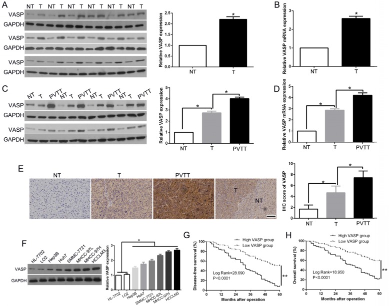
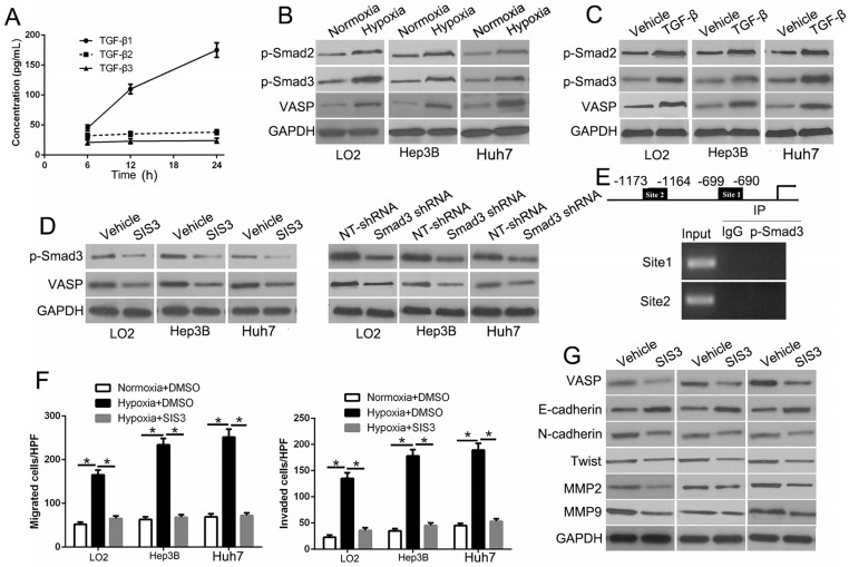
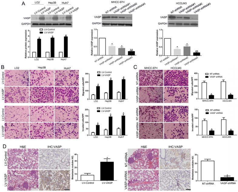
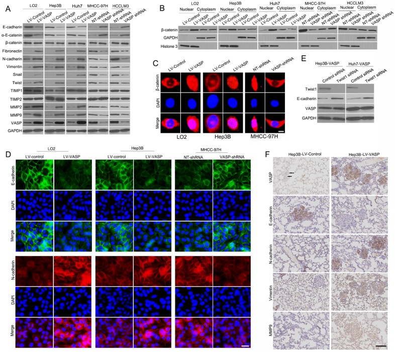
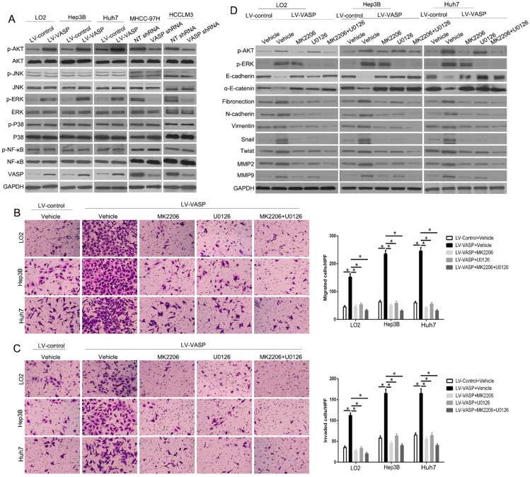
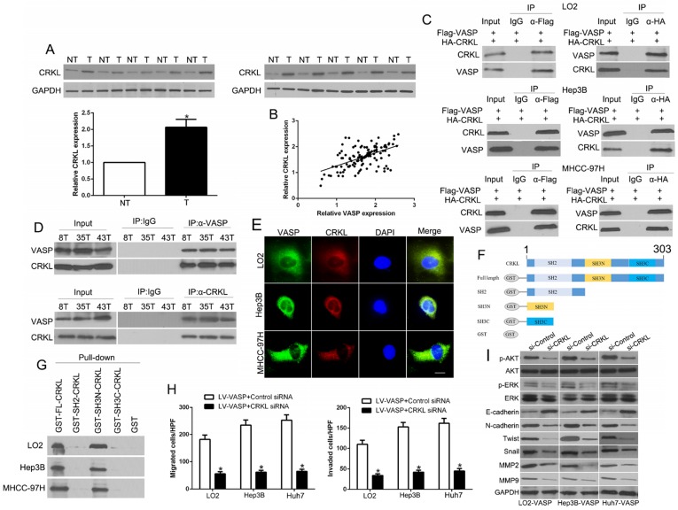
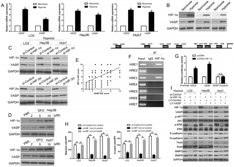
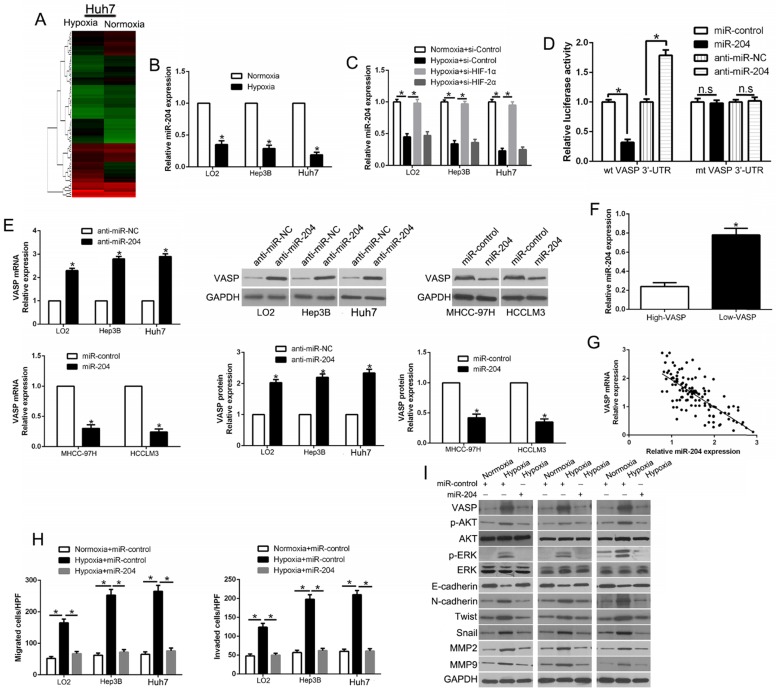
Patients with hepatocellular carcinoma (HCC) have a poor prognosis mostly due to intrahepatic as well as distal metastasis. Vasodilator-stimulated phosphoprotein (VASP), a regulator of actin cytoskeleton and cell migration, is overexpressed in HCC and correlated with its malignant features and poor prognosis. Very little is known about its function in HCC. qRT-PCR, Western blot and IHC were used to detect the VASP expression in tissues and cells. Transwell and wound healing assays were used to measure the migration and invasion of HCC cells. Immunoblotting and immunofluorescence were used for detection of epithelial-to-mesenchymal transition (EMT) progression in HCC cells. A lung metastasis mouse model was used to evaluate metastasis of HCC in vivo. The putative targets of miR-204 were disclosed by public databases and a dual-luciferase reporter assay. IP was used to show the interaction between VASP and CRKL. ChIP was used to analyze the binding of HIF-1α to VASP promoter region. Our data involving both gain- and loss-of-function studies revealed that VASP activated AKT and ERK signaling and promoted HCC migration and invasion in vitro and in vivo by altering the EMT phenotype and expression of MMPs. We investigated the positive correlation between VASP and an adapter protein, CRKL. VASP dynamically co-localized at the SH3N domain of CRKL and mediated its function. Mechanistically, VASP overexpression at the transcriptional level was mediated by HIF-1α through direct binding to two hypoxia response elements (HRE) in the VASP promoter region. Furthermore, we identified hypoxia-induced down-regulation of miR-204, which functioned as the regulator of VASP overexpression at the post-transcriptional level. Also, hypoxia-activated p-Smad3 dependent TGF-β signaling indirectly promoted VASP expression. A variety of hypoxia-induced molecular mechanisms contributed to the upregulation of VASP at transcriptional and post-transcriptional levels. These mechanisms involved CRKL, HIF-1α, miR-204, and TGF-β activating the AKT and ERK signaling to promote EMT and expression of MMPs. Taken together, our results defined VASP as an oncogene of HCC pathogenesis and metastasis with the potential to serve as a prognostic biomarker.
肝细胞癌(HCC)患者的预后较差,主要是由于肝内和远处转移。血管扩张刺激磷蛋白(VASP)是肌动蛋白细胞骨架和细胞迁移的调节剂,在 HCC 中过度表达,与恶性特征和不良预后相关。关于其在 HCC 中的功能知之甚少。qRT-PCR、Western blot 和 IHC 用于检测组织和细胞中的 VASP 表达。Transwell 和划痕愈合试验用于测量 HCC 细胞的迁移和侵袭。免疫印迹和免疫荧光用于检测 HCC 细胞上皮间质转化(EMT)的进展。肺转移小鼠模型用于评估 HCC 在体内的转移。通过公共数据库和双荧光素酶报告基因检测揭示了 miR-204 的假定靶标。IP 用于显示 VASP 和 CRKL 之间的相互作用。ChIP 用于分析 HIF-1α 与 VASP 启动子区域的结合。涉及增益和失能研究的数据表明,VASP 通过改变 EMT 表型和 MMPs 的表达,激活 AKT 和 ERK 信号通路,促进 HCC 在体外和体内的迁移和侵袭。我们研究了 VASP 与衔接蛋白 CRKL 之间的正相关关系。VASP 在 CRKL 的 SH3N 结构域动态共定位,并介导其功能。从机制上讲,转录水平上的 VASP 过表达是由 HIF-1α 通过直接结合 VASP 启动子区域的两个缺氧反应元件(HRE)介导的。此外,我们鉴定了缺氧诱导的 miR-204 下调,其在转录后水平作为 VASP 过表达的调节剂。此外,缺氧激活的 p-Smad3 依赖性 TGF-β 信号间接促进了 VASP 的表达。多种缺氧诱导的分子机制导致 VASP 在转录和转录后水平的上调。这些机制涉及 CRKL、HIF-1α、miR-204 和 TGF-β 激活 AKT 和 ERK 信号通路,促进 EMT 和 MMPs 的表达。总之,我们的研究结果将 VASP 定义为 HCC 发病机制和转移的癌基因,具有作为预后生物标志物的潜力。