Department of Internal Medicine, Krefting Research Centre, Institute of Medicine, The Sahlgrenska Academy, University of Gothenburg, Gothenburg, Sweden.
PLoS One. 2011;6(5):e19889. doi: 10.1371/journal.pone.0019889. Epub 2011 May 19.
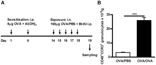
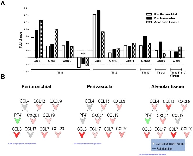
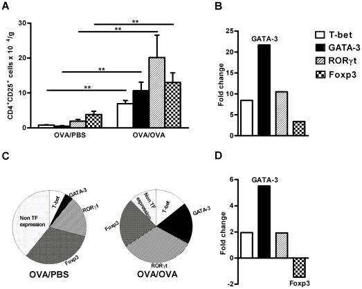
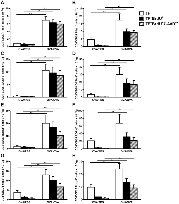
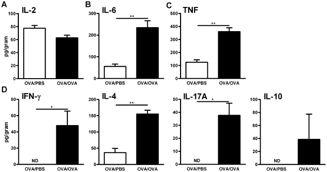
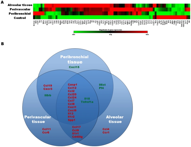
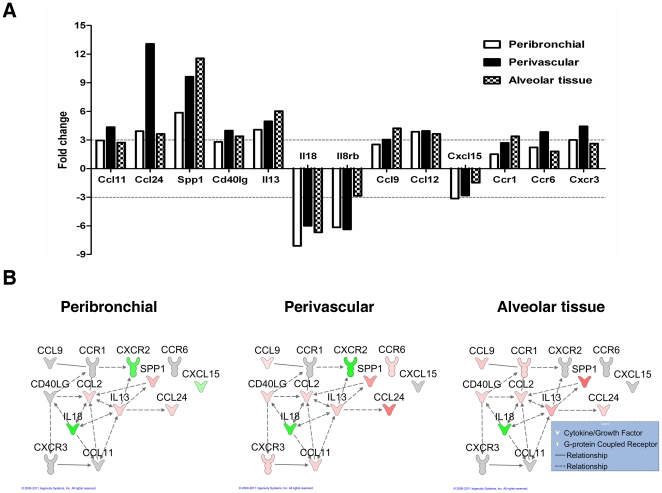
Allergic asthma is associated with airway eosinophilia, which is regulated by different T-effector cells. T cells express transcription factors T-bet, GATA-3, RORγt and Foxp3, representing Th1, Th2, Th17 and Treg cells respectively. No study has directly determined the relative presence of each of these T cell subsets concomitantly in a model of allergic airway inflammation. In this study we determined the degree of expansion of these T cell subsets, in the lungs of allergen challenged mice. Cell proliferation was determined by incorporation of 5-bromo-2'-deoxyuridine (BrdU) together with 7-aminoactnomycin (7-AAD). The immunohistochemical localisation of T cells in the lung microenvironments was also quantified. Local expression of cytokines, chemokines and receptor genes was measured using real-time RT-PCR array analysis in tissue sections isolated by laser microdissection and pressure catapulting technology. Allergen exposure increased the numbers of T-bet(+), GATA-3(+), RORγt(+) and Foxp3(+) cells in CD4(+)CD25(+) and CD4(+)CD25(-) T cells, with the greatest expansion of GATA-3(+) cells. The majority of CD4(+)CD25(+) T-bet(+), GATA-3(+), RORγt(+) and Foxp3(+) cells had incorporated BrdU and underwent proliferation during allergen exposure. Allergen exposure led to the accumulation of T-bet(+), GATA-3(+) and Foxp3(+) cells in peribronchial and alveolar tissue, GATA-3(+) and Foxp3(+) cells in perivascular tissue, and RORγt(+) cells in alveolar tissue. A total of 28 cytokines, chemokines and receptor genes were altered more than 3 fold upon allergen exposure, with expression of half of the genes claimed in all three microenvironments. Our study shows that allergen exposure affects all T effector cells in lung, with a dominant of Th2 cells, but with different local cell distribution, probably due to a distinguished local inflammatory milieu.
变应性哮喘与气道嗜酸性粒细胞增多有关,后者受不同的 T 效应细胞调节。T 细胞表达转录因子 T-bet、GATA-3、RORγt 和 Foxp3,分别代表 Th1、Th2、Th17 和 Treg 细胞。尚无研究直接确定这些 T 细胞亚群在变应性气道炎症模型中同时存在的相对程度。在这项研究中,我们确定了在过敏原挑战的小鼠肺部这些 T 细胞亚群的扩增程度。通过将 5-溴-2'-脱氧尿苷 (BrdU) 与 7-氨基放线菌素 (7-AAD) 一起掺入来确定细胞增殖。还通过激光微切割和压力弹射技术分离组织切片,使用实时 RT-PCR 阵列分析来定量测量肺微环境中 T 细胞的免疫组织化学定位。局部表达的细胞因子、趋化因子和受体基因使用实时 RT-PCR 阵列分析在激光微切割和压力弹射技术分离的组织切片中进行测量。过敏原暴露增加了 CD4(+)CD25(+) 和 CD4(+)CD25(-)T 细胞中 T-bet(+)、GATA-3(+)、RORγt(+) 和 Foxp3(+)细胞的数量,其中 GATA-3(+)细胞的扩增最大。在过敏原暴露期间,大多数 CD4(+)CD25(+)T-bet(+)、GATA-3(+)、RORγt(+) 和 Foxp3(+)细胞已掺入 BrdU 并经历增殖。过敏原暴露导致 T-bet(+)、GATA-3(+) 和 Foxp3(+)细胞在细支气管和肺泡组织中积聚,GATA-3(+)和 Foxp3(+)细胞在血管周围组织中积聚,RORγt(+)细胞在肺泡组织中积聚。过敏原暴露后共有 28 种细胞因子、趋化因子和受体基因的表达增加了 3 倍以上,其中一半的基因在所有三种微环境中都有表达。我们的研究表明,过敏原暴露会影响肺部的所有 T 效应细胞,以 Th2 细胞为主,但具有不同的局部细胞分布,可能是由于独特的局部炎症环境所致。