Department of Neurosurgery, Medical College of Wisconsin, 8701 Watertown Plank Road, Milwaukee, WI 53226, USA.
Brain Res. 2011 Jul 5;1399:15-24. doi: 10.1016/j.brainres.2011.05.018. Epub 2011 May 15.
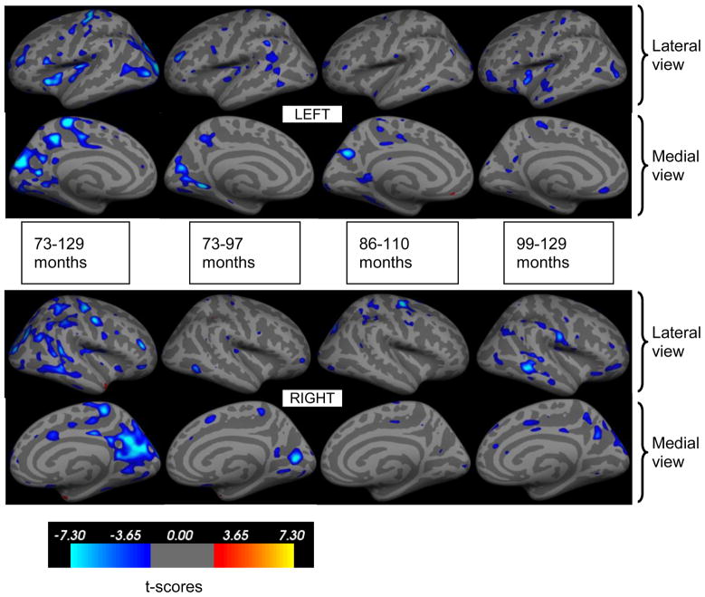
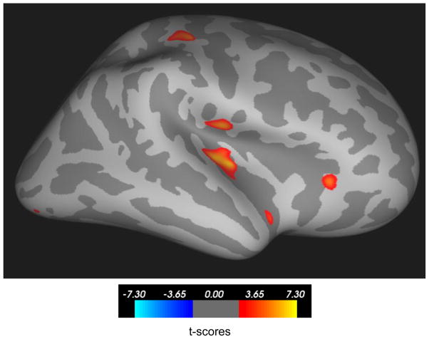
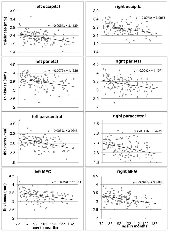
There is evidence that abnormal cerebral development during childhood is a risk factor for various cognitive and psychiatric disorders. There is not, however, sufficient normative data available on large samples of typically developing children, especially within the narrow preadolescent age range. We analyzed high resolution MRI images from 126 normally developing children between ages 6 and 10 years. Age related differences in cortical thickness and in the volumes of major subcortical structures were assessed. Thinner cortices were observed in the occipital, parietal and somatosensory regions as well as in distinct regions of the temporal and frontal lobes with increasing age. Among the major subcortical structures analyzed in this study, only the thalamus showed increased volume with age after accounting for intracranial volume. Within the age range studied age-related cortical and subcortical differences were similar for boys and girls except for the right insula, where girls showed a slight increase in thickness with age. The findings reveal age-associated changes in brain anatomy, providing information about the trajectory of normal brain development during late childhood.
有证据表明,儿童期大脑发育异常是各种认知和精神障碍的一个风险因素。然而,在正常发育儿童的大样本中,特别是在狭窄的青春期前年龄段,没有足够的规范数据。我们分析了年龄在 6 至 10 岁之间的 126 名正常发育儿童的高分辨率 MRI 图像。评估了皮质厚度和主要皮质下结构体积随年龄的变化。随着年龄的增长,观察到枕叶、顶叶和体感区域以及颞叶和额叶的特定区域的皮质变薄。在本研究分析的主要皮质下结构中,只有丘脑在考虑到颅内体积后,随着年龄的增长而体积增加。在所研究的年龄范围内,男孩和女孩的皮质和皮质下差异与年龄相关,除了右侧脑岛,女孩的脑岛厚度随年龄略有增加。这些发现揭示了大脑解剖结构与年龄相关的变化,为了解儿童晚期正常大脑发育的轨迹提供了信息。